5HV4

Crystal Structure of a Prolyl 4-Hydroxylase Complexed with Alpha-ketoglutarate from the Pathogenic Bacterium Bacillus anthracis in C2221


Experimental Data Snapshot

  • Method: X-RAY DIFFRACTION
  • Resolution: 2.35 Å
  • R-Value Free: 0.245 
  • R-Value Work: 0.199 
  • R-Value Observed: 0.201 

wwPDB Validation   3D Report Full Report


This is version 1.2 of the entry. See complete history


Literature

Structural analysis of cofactor binding for a prolyl 4-hydroxylase from the pathogenic bacterium Bacillus anthracis.

Schnicker, N.J.Dey, M.

(2016) Acta Crystallogr D Struct Biol 72: 675-681

  • DOI: https://doi.org/10.1107/S2059798316004198
  • Primary Citation of Related Structures:  
    5HV0, 5HV4

  • PubMed Abstract: 

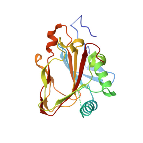
    The prolyl 4-hydroxylases (P4Hs) are mononuclear nonheme iron enzymes that catalyze the formation of 4R-hydroxyproline from many different substrates, with various biological implications. P4H is a key player in collagen accumulation, which has implications in fibrotic disorders. The stabilization of collagen triple-helical structure via prolyl hydroxylation is the rate-limiting step in collagen biosynthesis, and therefore P4H has been extensively investigated as a potential therapeutic target of fibrotic disease. Understanding how these enzymes recognize cofactors and substrates is important and will aid in the future design of inhibitors of P4H. In this article, X-ray crystal structures of a metallocofactor- and α-ketoglutarate (αKG)-bound form of P4H from Bacillus anthracis (BaP4H) are reported. Structures of BaP4H were solved at 1.63 and 2.35 Å resolution and contained a cadmium ion and αKG bound in the active site. The αKG-Cd-BaP4H ternary complex reveals conformational changes of conserved residues upon the binding of metal ion and αKG, resulting in a closed active-site configuration required for dioxygen, substrate binding and catalysis.


  • Organizational Affiliation

    Department of Chemistry, University of Iowa, Iowa City, IA 52242, USA.


Macromolecules
Find similar proteins by:  (by identity cutoff)  |  3D Structure
Entity ID: 1
MoleculeChains Sequence LengthOrganismDetailsImage
2OG-Fe(II) oxygenase217Bacillus anthracisMutation(s): 0 
Gene Names: 
UniProt
Find proteins for A0A4Y1WAP5 (Bacillus anthracis)
Explore A0A4Y1WAP5 
Go to UniProtKB:  A0A4Y1WAP5
Entity Groups  
Sequence Clusters30% Identity50% Identity70% Identity90% Identity95% Identity100% Identity
UniProt GroupA0A4Y1WAP5
Sequence Annotations
Expand
  • Reference Sequence
Small Molecules
Ligands 3 Unique
IDChains Name / Formula / InChI Key2D Diagram3D Interactions
AKG
Query on AKG

Download Ideal Coordinates CCD File 
G [auth A]2-OXOGLUTARIC ACID
C5 H6 O5
KPGXRSRHYNQIFN-UHFFFAOYSA-N
CD
Query on CD

Download Ideal Coordinates CCD File 
B [auth A],
C [auth A],
D [auth A],
E [auth A],
F [auth A]
CADMIUM ION
Cd
WLZRMCYVCSSEQC-UHFFFAOYSA-N
K
Query on K

Download Ideal Coordinates CCD File 
H [auth A]
I [auth A]
J [auth A]
K [auth A]
L [auth A]
H [auth A],
I [auth A],
J [auth A],
K [auth A],
L [auth A],
M [auth A]
POTASSIUM ION
K
NPYPAHLBTDXSSS-UHFFFAOYSA-N
Binding Affinity Annotations 
IDSourceBinding Affinity
AKG Binding MOAD:  5HV4 Kd: 1100 (nM) from 1 assay(s)
Experimental Data & Validation

Experimental Data

Unit Cell:
Length ( Å )Angle ( ˚ )
a = 42.551α = 90
b = 146.189β = 90
c = 75.075γ = 90
Software Package:
Software NamePurpose
PHENIXrefinement
SCALAdata scaling
PDB_EXTRACTdata extraction
XDSdata reduction
PHASERphasing

Structure Validation

View Full Validation Report


Entry History & Funding Information

Deposition Data


Funding OrganizationLocationGrant Number
College of Liberal Arts and Sciences University of IowaUnited States--

Revision History  (Full details and data files)

  • Version 1.0: 2016-05-04
    Type: Initial release
  • Version 1.1: 2016-05-18
    Changes: Database references
  • Version 1.2: 2023-09-27
    Changes: Data collection, Database references, Derived calculations, Refinement description