5GW0

Crystal structure of SNX16 PX-Coiled coil


Experimental Data Snapshot

  • Method: X-RAY DIFFRACTION
  • Resolution: 3.30 Å
  • R-Value Free: 0.288 
  • R-Value Work: 0.236 
  • R-Value Observed: 0.239 

wwPDB Validation   3D Report Full Report


This is version 1.0 of the entry. See complete history


Literature

SNX16 Regulates the Recycling of E-Cadherin through a Unique Mechanism of Coordinated Membrane and Cargo Binding.

Xu, J.Zhang, L.Ye, Y.Shan, Y.Wan, C.Wang, J.Pei, D.Shu, X.Liu, J.

(2017) Structure 25: 1251-1263.e5

  • DOI: https://doi.org/10.1016/j.str.2017.06.015
  • Primary Citation of Related Structures:  
    5GW0, 5GW1

  • PubMed Abstract: 

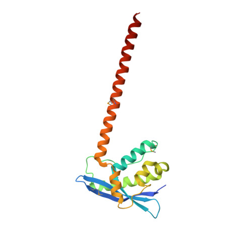
    E-Cadherin is a major component of adherens junctions on cell surfaces. SNX16 is a unique member of sorting nexins that contains a coiled-coil (CC) domain downstream of the PX domain. We report here that SNX16 regulates the recycling trafficking of E-cadherin. We solved the crystal structure of PX-CC unit of SNX16 and revealed a unique shear shaped homodimer. We identified a novel PI3P binding pocket in SNX16 that consists of both the PX and the CC domains. Surprisingly, we showed that the PPII/α2 loop, which is generally regarded as a membrane insertion loop in PX family proteins, is involved in the E-cadherin binding with SNX16. We then proposed a multivalent membrane binding model for SNX16. Our study postulates a new mechanism for coordinated membrane binding and cargo binding for SNX family proteins in general, and provide novel insights into recycling trafficking of E-cadherin.


  • Organizational Affiliation

    State Key Laboratory of Respiratory Disease, Guangzhou Institutes of Biomedicine and Health, Chinese Academy of Sciences, Guangzhou 510530, China.


Macromolecules
Find similar proteins by:  (by identity cutoff)  |  3D Structure
Entity ID: 1
MoleculeChains Sequence LengthOrganismDetailsImage
Sorting nexin-16
A, B, C, D, E
A, B, C, D, E, F
179Homo sapiensMutation(s): 0 
Gene Names: SNX16
UniProt & NIH Common Fund Data Resources
Find proteins for P57768 (Homo sapiens)
Explore P57768 
Go to UniProtKB:  P57768
PHAROS:  P57768
GTEx:  ENSG00000104497 
Entity Groups  
Sequence Clusters30% Identity50% Identity70% Identity90% Identity95% Identity100% Identity
UniProt GroupP57768
Sequence Annotations
Expand
  • Reference Sequence
Small Molecules
Modified Residues  1 Unique
IDChains TypeFormula2D DiagramParent
MSE
Query on MSE
A, B, C, D, E
A, B, C, D, E, F
L-PEPTIDE LINKINGC5 H11 N O2 SeMET
Experimental Data & Validation

Experimental Data

  • Method: X-RAY DIFFRACTION
  • Resolution: 3.30 Å
  • R-Value Free: 0.288 
  • R-Value Work: 0.236 
  • R-Value Observed: 0.239 
  • Space Group: P 1 21 1
Unit Cell:
Length ( Å )Angle ( ˚ )
a = 70.38α = 90
b = 196.78β = 111.75
c = 72.96γ = 90
Software Package:
Software NamePurpose
PHENIXrefinement
MOSFLMdata collection
Aimlessdata scaling
PDB_EXTRACTdata extraction
Aimlessdata reduction
MR-Rosettaphasing

Structure Validation

View Full Validation Report


Entry History 

Deposition Data

  • Released Date: 2017-09-13 
  • Deposition Author(s): Xu, J., Liu, J.

Revision History  (Full details and data files)

  • Version 1.0: 2017-09-13
    Type: Initial release