5CVY

The Structure of Bacillus pumilus GH48 in complex with cellobiose and cellohexaose


Experimental Data Snapshot

  • Method: X-RAY DIFFRACTION
  • Resolution: 2.00 Å
  • R-Value Free: 0.189 
  • R-Value Work: 0.146 
  • R-Value Observed: 0.148 

wwPDB Validation   3D Report Full Report


This is version 2.1 of the entry. See complete history


Literature

Natural diversity of glycoside hydrolase family 48 exoglucanases: insights from structure.

Brunecky, R.Alahuhta, M.Sammond, D.W.Xu, Q.Chen, M.Wilson, D.B.Brady, J.W.Himmel, M.E.Bomble, Y.J.Lunin, V.V.

(2017) Biotechnol Biofuels 10: 274-274

  • DOI: https://doi.org/10.1186/s13068-017-0951-5
  • Primary Citation of Related Structures:  
    5CVY

  • PubMed Abstract: 

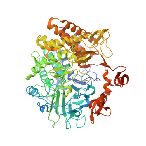
    Glycoside hydrolase (GH) family 48 is an understudied and increasingly important exoglucanase family found in the majority of bacterial cellulase systems. Moreover, many thermophilic enzyme systems contain GH48 enzymes. Deletion of GH48 enzymes in these microorganisms results in drastic reduction in biomass deconstruction. Surprisingly, given their importance for these microorganisms, GH48s have intrinsically low cellulolytic activity but even in low ratios synergize greatly with GH9 endoglucanases. In this study, we explore the structural and enzymatic diversity of these enzymes across a wide range of temperature optima. We have crystallized one new GH48 module from Bacillus pumilus in a complex with cellobiose and cellohexaose ( Bpum GH48). We compare this structure to other known GH48 enzymes in an attempt to understand GH48 structure/function relationships and draw general rules correlating amino acid sequences and secondary structures to thermostability in this GH family.


  • Organizational Affiliation

    Biosciences Center, National Renewable Energy Laboratory, 15013 Denver West Parkway, Golden, CO 80401 USA.


Macromolecules
Find similar proteins by:  (by identity cutoff)  |  3D Structure
Entity ID: 1
MoleculeChains Sequence LengthOrganismDetailsImage
Glycoside hydrolase709Bacillus pumilus SAFR-032Mutation(s): 0 
Gene Names: BPUM_1559
UniProt
Find proteins for A8FDC4 (Bacillus pumilus (strain SAFR-032))
Explore A8FDC4 
Go to UniProtKB:  A8FDC4
Entity Groups  
Sequence Clusters30% Identity50% Identity70% Identity90% Identity95% Identity100% Identity
UniProt GroupA8FDC4
Sequence Annotations
Expand
  • Reference Sequence
Oligosaccharides

Help

Entity ID: 2
MoleculeChains Length2D Diagram Glycosylation3D Interactions
beta-D-glucopyranose-(1-4)-beta-D-glucopyranose
B
2N/A
Glycosylation Resources
GlyTouCan:  G84824ZO
GlyCosmos:  G84824ZO
Entity ID: 3
MoleculeChains Length2D Diagram Glycosylation3D Interactions
beta-D-glucopyranose-(1-4)-beta-D-glucopyranose-(1-4)-beta-D-glucopyranose-(1-4)-beta-D-glucopyranose-(1-4)-beta-D-glucopyranose-(1-4)-beta-D-glucopyranose
C
6N/A
Glycosylation Resources
GlyTouCan:  G09454VW
GlyCosmos:  G09454VW
GlyGen:  G09454VW
Small Molecules
Ligands 5 Unique
IDChains Name / Formula / InChI Key2D Diagram3D Interactions
MLI
Query on MLI

Download Ideal Coordinates CCD File 
E [auth A],
F [auth A],
G [auth A]
MALONATE ION
C3 H2 O4
OFOBLEOULBTSOW-UHFFFAOYSA-L
GOL
Query on GOL

Download Ideal Coordinates CCD File 
V [auth A],
W [auth A],
X [auth A],
Y [auth A]
GLYCEROL
C3 H8 O3
PEDCQBHIVMGVHV-UHFFFAOYSA-N
EDO
Query on EDO

Download Ideal Coordinates CCD File 
H [auth A]
I [auth A]
J [auth A]
K [auth A]
L [auth A]
H [auth A],
I [auth A],
J [auth A],
K [auth A],
L [auth A],
M [auth A],
N [auth A],
O [auth A],
P [auth A],
Q [auth A],
R [auth A],
S [auth A],
T [auth A],
U [auth A]
1,2-ETHANEDIOL
C2 H6 O2
LYCAIKOWRPUZTN-UHFFFAOYSA-N
CA
Query on CA

Download Ideal Coordinates CCD File 
D [auth A]CALCIUM ION
Ca
BHPQYMZQTOCNFJ-UHFFFAOYSA-N
NA
Query on NA

Download Ideal Coordinates CCD File 
AA [auth A]
BA [auth A]
CA [auth A]
DA [auth A]
EA [auth A]
AA [auth A],
BA [auth A],
CA [auth A],
DA [auth A],
EA [auth A],
FA [auth A],
GA [auth A],
HA [auth A],
IA [auth A],
JA [auth A],
KA [auth A],
LA [auth A],
MA [auth A],
NA [auth A],
Z [auth A]
SODIUM ION
Na
FKNQFGJONOIPTF-UHFFFAOYSA-N
Biologically Interesting Molecules (External Reference) 2 Unique
Experimental Data & Validation

Experimental Data

  • Method: X-RAY DIFFRACTION
  • Resolution: 2.00 Å
  • R-Value Free: 0.189 
  • R-Value Work: 0.146 
  • R-Value Observed: 0.148 
  • Space Group: P 43 21 2
Unit Cell:
Length ( Å )Angle ( ˚ )
a = 98.041α = 90
b = 98.041β = 90
c = 218.956γ = 90
Software Package:
Software NamePurpose
REFMACrefinement
MOLREPphasing
Cootmodel building

Structure Validation

View Full Validation Report


Entry History 

Deposition Data

Revision History  (Full details and data files)

  • Version 1.0: 2016-08-10
    Type: Initial release
  • Version 1.1: 2019-06-05
    Changes: Data collection, Database references, Derived calculations
  • Version 2.0: 2020-07-29
    Type: Remediation
    Reason: Carbohydrate remediation
    Changes: Atomic model, Data collection, Derived calculations, Non-polymer description, Structure summary
  • Version 2.1: 2023-09-27
    Changes: Data collection, Database references, Refinement description, Structure summary