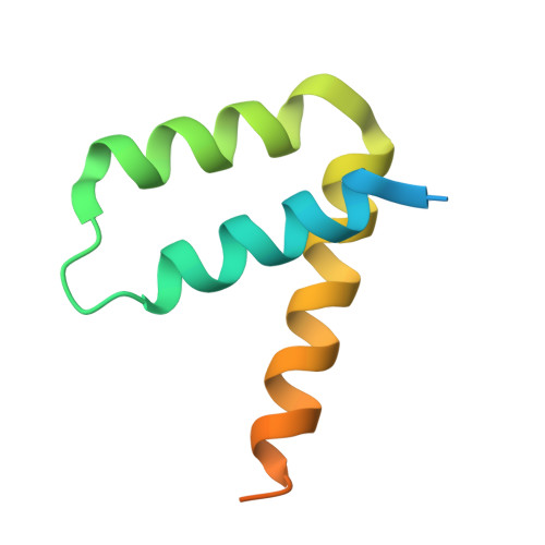
Mechanistic Implications of the Unique Structural Features and Dimerization of the Cytoplasmic Domain of the Pseudomonas Sigma Regulator, PupR.
Jensen, J.L., Balbo, A., Neau, D.B., Chakravarthy, S., Zhao, H., Sinha, S.C., Colbert, C.L.(2015) Biochemistry 54: 5867-5877
- PubMed: 26313375
- DOI: https://doi.org/10.1021/acs.biochem.5b00826
- Primary Citation of Related Structures:
5CAM, 5COS - PubMed Abstract:
Gram-negative bacteria tightly regulate intracellular levels of iron, an essential nutrient. To ensure this strict control, some outer membrane TonB-dependent transporters (TBDTs) that are responsible for iron import stimulate their own transcription in response to extracellular binding by an iron-laden siderophore. This process is mediated by an inner membrane sigma regulator protein (an anti-sigma factor) that transduces an unknown periplasmic signal from the TBDT to release an intracellular sigma factor from the inner membrane, which ultimately upregulates TBDT transcription. Here, we use the Pseudomonas putida ferric-pseudobactin BN7/BN8 sigma regulator, PupR, as a model system to understand the molecular mechanism of this conserved class of sigma regulators. We have determined the X-ray crystal structure of the cytoplasmic anti-sigma domain (ASD) of PupR to 2.0 Å. Size exclusion chromatography, small-angle X-ray scattering, and sedimentation velocity analytical ultracentrifugation all indicate that, in contrast to other ASDs, the PupR-ASD exists as a dimer in solution. Mutagenesis of residues at the dimer interface identified from the crystal structure disrupts dimerization and protein stability, as determined by sedimentation velocity analytical ultracentrifugation and thermal denaturation circular dichroism spectroscopy. These combined results suggest that this type of inner membrane sigma regulator may utilize an unusual mechanism to sequester their cognate sigma factors and prevent transcription activation.
Organizational Affiliation:
Department of Chemistry and Biochemistry, North Dakota State University , Fargo, North Dakota 58108-6050, United States.