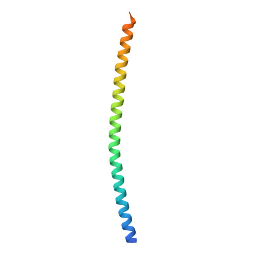
The structure of the GemC1 coiled coil and its interaction with the Geminin family of coiled-coil proteins.
Caillat, C., Fish, A., Pefani, D.E., Taraviras, S., Lygerou, Z., Perrakis, A.(2015) Acta Crystallogr D Biol Crystallogr 71: 2278-2286
- PubMed: 26527144
- DOI: https://doi.org/10.1107/S1399004715016892
- Primary Citation of Related Structures:
5C9N - PubMed Abstract:
GemC1, together with Idas and Geminin, an important regulator of DNA-replication licensing and differentiation decisions, constitute a superfamily sharing a homologous central coiled-coil domain. To better understand this family of proteins, the crystal structure of a GemC1 coiled-coil domain variant engineered for better solubility was determined to 2.2 Å resolution. GemC1 shows a less typical coiled coil compared with the Geminin homodimer and the Geminin-Idas heterodimer structures. It is also shown that both in vitro and in cells GemC1 interacts with Geminin through its coiled-coil domain, forming a heterodimer that is more stable that the GemC1 homodimer. Comparative analysis of the thermal stability of all of the possible superfamily complexes, using circular dichroism to follow the unfolding of the entire helix of the coiled coil, or intrinsic tryptophan fluorescence of a unique conserved N-terminal tryptophan, shows that the unfolding of the coiled coil is likely to take place from the C-terminus towards the N-terminus. It is also shown that homodimers show a single-state unfolding, while heterodimers show a two-state unfolding, suggesting that the dimer first falls apart and the helices then unfold according to the stability of each protein. The findings argue that Geminin-family members form homodimers and heterodimers between them, and this ability is likely to be important for modulating their function in cycling and differentiating cells.
Organizational Affiliation:
Department of Biochemistry, The Netherlands Cancer Institute, 1066 CX Amsterdam, The Netherlands.