5AOF

Crystal structure of pneumolysin deletion mutant Delta146_147.


Experimental Data Snapshot

  • Method: X-RAY DIFFRACTION
  • Resolution: 2.45 Å
  • R-Value Free: 0.234 
  • R-Value Work: 0.208 
  • R-Value Observed: 0.209 

wwPDB Validation   3D Report Full Report


This is version 1.2 of the entry. See complete history


Literature

CryoEM structures of membrane pore and prepore complex reveal cytolytic mechanism of Pneumolysin.

van Pee, K.Neuhaus, A.D'Imprima, E.Mills, D.J.Kuhlbrandt, W.Yildiz, O.

(2017) Elife 6

  • DOI: https://doi.org/10.7554/eLife.23644
  • Primary Citation of Related Structures:  
    5AOE, 5AOF, 5LY6

  • PubMed Abstract: 

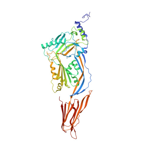
    Many pathogenic bacteria produce pore-forming toxins to attack and kill human cells. We have determined the 4.5 Å structure of the ~2.2 MDa pore complex of pneumolysin, the main virulence factor of Streptococcus pneumoniae , by cryoEM. The pneumolysin pore is a 400 Å ring of 42 membrane-inserted monomers. Domain 3 of the soluble toxin refolds into two ~85 Å β-hairpins that traverse the lipid bilayer and assemble into a 168-strand β-barrel. The pore complex is stabilized by salt bridges between β-hairpins of adjacent subunits and an internal α-barrel. The apolar outer barrel surface with large sidechains is immersed in the lipid bilayer, while the inner barrel surface is highly charged. Comparison of the cryoEM pore complex to the prepore structure obtained by electron cryo-tomography and the x-ray structure of the soluble form reveals the detailed mechanisms by which the toxin monomers insert into the lipid bilayer to perforate the target membrane.


  • Organizational Affiliation

    Department of Structural Biology, Max Planck Institute of Biophysics, Frankfurt am Main, Germany.


Macromolecules
Find similar proteins by:  (by identity cutoff)  |  3D Structure
Entity ID: 1
MoleculeChains Sequence LengthOrganismDetailsImage
PNEUMOLYSIN488Streptococcus pneumoniaeMutation(s): 0 
Membrane Entity: Yes 
UniProt
Find proteins for Q7ZAK5 (Streptococcus pneumoniae (strain ATCC BAA-255 / R6))
Explore Q7ZAK5 
Go to UniProtKB:  Q7ZAK5
Entity Groups  
Sequence Clusters30% Identity50% Identity70% Identity90% Identity95% Identity100% Identity
UniProt GroupQ7ZAK5
Sequence Annotations
Expand
  • Reference Sequence
Experimental Data & Validation

Experimental Data

  • Method: X-RAY DIFFRACTION
  • Resolution: 2.45 Å
  • R-Value Free: 0.234 
  • R-Value Work: 0.208 
  • R-Value Observed: 0.209 
  • Space Group: P 21 21 21
Unit Cell:
Length ( Å )Angle ( ˚ )
a = 24.73α = 90
b = 163.5β = 90
c = 207.8γ = 90
Software Package:
Software NamePurpose
PHENIXrefinement
XDSdata reduction
XSCALEdata scaling
PHASERphasing

Structure Validation

View Full Validation Report


Entry History 

Deposition Data

Revision History  (Full details and data files)

  • Version 1.0: 2016-10-05
    Type: Initial release
  • Version 1.1: 2017-04-05
    Changes: Database references
  • Version 1.2: 2024-01-10
    Changes: Data collection, Database references, Other, Refinement description