5AJI

MscS D67R1 high resolution


Experimental Data Snapshot

  • Method: X-RAY DIFFRACTION
  • Resolution: 2.99 Å
  • R-Value Free: 0.263 
  • R-Value Work: 0.244 
  • R-Value Observed: 0.245 

wwPDB Validation   3D Report Full Report


This is version 1.3 of the entry. See complete history


Literature

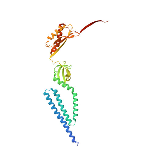
The Role of Lipids in Mechanosensation.

Pliotas, C.Dahl, A.C.E.Rasmussen, T.Mahendran, K.R.Smith, T.K.Marius, P.Gault, J.Banda, T.Rasmussen, A.Miller, S.Robinson, C.V.Bayley, H.Sansom, M.S.P.Booth, I.R.Naismith, J.H.

(2015) Nat Struct Mol Biol 22: 991

  • DOI: https://doi.org/10.1038/nsmb.3120
  • Primary Citation of Related Structures:  
    5AJI

  • PubMed Abstract: 

    The ability of proteins to sense membrane tension is pervasive in biology. A higher-resolution structure of the Escherichia coli small-conductance mechanosensitive channel MscS identifies alkyl chains inside pockets formed by the transmembrane helices (TMs). Purified MscS contains E. coli lipids, and fluorescence quenching demonstrates that phospholipid acyl chains exchange between bilayer and TM pockets. Molecular dynamics and biophysical analyses show that the volume of the pockets and thus the number of lipid acyl chains within them decreases upon channel opening. Phospholipids with one acyl chain per head group (lysolipids) displace normal phospholipids (with two acyl chains) from MscS pockets and trigger channel opening. We propose that the extent of acyl-chain interdigitation in these pockets determines the conformation of MscS. When interdigitation is perturbed by increased membrane tension or by lysolipids, the closed state becomes unstable, and the channel gates.


  • Organizational Affiliation

    Biomedical Sciences Research Complex, University of St. Andrews, St. Andrews, UK.


Macromolecules
Find similar proteins by:  (by identity cutoff)  |  3D Structure
Entity ID: 1
MoleculeChains Sequence LengthOrganismDetailsImage
SMALL-CONDUCTANCE MECHANOSENSITIVE CHANNEL
A, B, C, D, E
A, B, C, D, E, F, G
286Escherichia coliMutation(s): 1 
Membrane Entity: Yes 
UniProt
Find proteins for P0C0S1 (Escherichia coli (strain K12))
Explore P0C0S1 
Go to UniProtKB:  P0C0S1
Entity Groups  
Sequence Clusters30% Identity50% Identity70% Identity90% Identity95% Identity100% Identity
UniProt GroupP0C0S1
Sequence Annotations
Expand
  • Reference Sequence
Small Molecules
Ligands 3 Unique
IDChains Name / Formula / InChI Key2D Diagram3D Interactions
D10
Query on D10

Download Ideal Coordinates CCD File 
L [auth C],
O [auth D],
V [auth G]
DECANE
C10 H22
DIOQZVSQGTUSAI-UHFFFAOYSA-N
OCT
Query on OCT

Download Ideal Coordinates CCD File 
H [auth A],
Q [auth E]
N-OCTANE
C8 H18
TVMXDCGIABBOFY-UHFFFAOYSA-N
HEX
Query on HEX

Download Ideal Coordinates CCD File 
I [auth A]
J [auth B]
K [auth B]
M [auth C]
N [auth C]
I [auth A],
J [auth B],
K [auth B],
M [auth C],
N [auth C],
P [auth D],
R [auth E],
S [auth E],
T [auth F],
U [auth F]
HEXANE
C6 H14
VLKZOEOYAKHREP-UHFFFAOYSA-N
Modified Residues  1 Unique
IDChains TypeFormula2D DiagramParent
R1A
Query on R1A
A, B, C, D, E
A, B, C, D, E, F, G
L-PEPTIDE LINKINGC12 H21 N2 O3 S2CYS
Experimental Data & Validation

Experimental Data

  • Method: X-RAY DIFFRACTION
  • Resolution: 2.99 Å
  • R-Value Free: 0.263 
  • R-Value Work: 0.244 
  • R-Value Observed: 0.245 
  • Space Group: P 21 21 21
Unit Cell:
Length ( Å )Angle ( ˚ )
a = 126.33α = 90
b = 149.09β = 90
c = 173.82γ = 90
Software Package:
Software NamePurpose
REFMACrefinement

Structure Validation

View Full Validation Report


Entry History 

Deposition Data

Revision History  (Full details and data files)

  • Version 1.0: 2015-07-08
    Type: Initial release
  • Version 1.1: 2015-11-18
    Changes: Database references
  • Version 1.2: 2015-12-23
    Changes: Database references
  • Version 1.3: 2024-01-10
    Changes: Data collection, Database references, Derived calculations, Other, Refinement description