5YSC

Crystal Structure of periplasmic Vitamin B12 binding protein BtuF of Vibrio cholerae


Experimental Data Snapshot

  • Method: X-RAY DIFFRACTION
  • Resolution: 1.67 Å
  • R-Value Free: 0.227 
  • R-Value Work: 0.197 
  • R-Value Observed: 0.199 

wwPDB Validation   3D Report Full Report


Ligand Structure Quality Assessment 


This is version 2.1 of the entry. See complete history


Literature

Mechanistic basis of vitamin B12 and cobinamide salvaging by the Vibrio species.

Agarwal, S.Dey, S.Ghosh, B.Biswas, M.Dasgupta, J.

(2019) Biochim Biophys Acta Proteins Proteom 1867: 140-151

  • DOI: https://doi.org/10.1016/j.bbapap.2018.11.004
  • Primary Citation of Related Structures:  
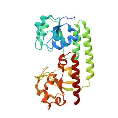
    5YSC

  • PubMed Abstract: 

    Biosynthesis of vitamin B12, which occurs through salvaging pathway or de novo synthesis, is essential for the survival and growth of bacteria. While the mechanism is known for many bacteria, it is elusive yet for diarrhea causing pathogenic bacteria Vibrio cholerae or the other Vibrio species. Sequence analysis using genome databases delineated that majority of the Vibrio species including V. cholerae contain genes required for salvaging cobalamin/cobinamide in aerobic pathway while lack the genes required for de novo synthesis of B12. Fluorescence quenching study showed that VcBtuF, the PBP of putative ABC transporter BtuF-CD of V. cholerae O395 binds cyanocobalamin and dicyanocobinamide with micromolar dissociation constants (K d ). Productive internalization of these nutrients has been established through growth assay. The crystal structure of cyanocobalamin bound VcBtuF has shown that although interactions between cyanocobalamin and VcBtuF are largely similar to E. coli BtuF, VcBtuF possesses a wider binding pocket. MD simulations indicated that in contrast to EcBtuF that executes 'open-close' movement, inter-lobe twisting is prevalent in VcBtuF. Although H70, located at the entrance of the substrate binding cleft of VcBtuF, executes swinging motion, it cannot act as 'closed gate' to retain cyanocobalamin or cobinamide in the pocket like corresponding residue W66 of EcBtuF. Rather, VcBtuF shows a distinctive phenomenon of heme binding with comparable affinity to B12. Soret shift of heme upon binding with VcBtuF pointed towards involvement of H70 in heme recognition. This may lead to a restricted B12 or cobinamide binding during abundance of heme in the periplasmic space.


  • Organizational Affiliation

    Department of Biotechnology, St. Xavier's College, 30 Park Street, Kolkata 700016, India.


Macromolecules
Find similar proteins by:  (by identity cutoff)  |  3D Structure
Entity ID: 1
MoleculeChains Sequence LengthOrganismDetailsImage
Vitamin B12-binding protein271Vibrio cholerae O395Mutation(s): 0 
Gene Names: btuF
UniProt
Find proteins for A5F5P5 (Vibrio cholerae serotype O1 (strain ATCC 39541 / Classical Ogawa 395 / O395))
Explore A5F5P5 
Go to UniProtKB:  A5F5P5
Entity Groups  
Sequence Clusters30% Identity50% Identity70% Identity90% Identity95% Identity100% Identity
UniProt GroupA5F5P5
Sequence Annotations
Expand
  • Reference Sequence
Small Molecules
Ligands 2 Unique
IDChains Name / Formula / InChI Key2D Diagram3D Interactions
CNC
Query on CNC

Download Ideal Coordinates CCD File 
B [auth A]CYANOCOBALAMIN
C63 H89 Co N14 O14 P
SYZBZQWSWIJYAR-UVKKECPRSA-M
SO4
Query on SO4

Download Ideal Coordinates CCD File 
C [auth A]SULFATE ION
O4 S
QAOWNCQODCNURD-UHFFFAOYSA-L
Experimental Data & Validation

Experimental Data

  • Method: X-RAY DIFFRACTION
  • Resolution: 1.67 Å
  • R-Value Free: 0.227 
  • R-Value Work: 0.197 
  • R-Value Observed: 0.199 
  • Space Group: P 65 2 2
Unit Cell:
Length ( Å )Angle ( ˚ )
a = 55.877α = 90
b = 55.877β = 90
c = 279.163γ = 120
Software Package:
Software NamePurpose
PHENIXrefinement
XDSdata reduction
SCALAdata scaling
PHASERphasing

Structure Validation

View Full Validation Report


Ligand Structure Quality Assessment 


Entry History 

Deposition Data

Revision History  (Full details and data files)

  • Version 1.0: 2018-09-19
    Type: Initial release
  • Version 1.1: 2019-04-17
    Changes: Data collection, Database references
  • Version 2.0: 2021-08-25
    Changes: Data collection, Database references, Derived calculations, Non-polymer description, Structure summary
  • Version 2.1: 2023-11-22
    Changes: Data collection, Refinement description