


Crystal Structure of L-Threonine Aldolase from Pseudomonas putida
Beaudoin, S.F., Burg, M.J., Stewart, J.D.To be published.
Experimental Data Snapshot
Entity ID: 1 | |||||
|---|---|---|---|---|---|
| Molecule | Chains | Sequence Length | Organism | Details | Image |
| L-threonine aldolase | 346 | Pseudomonas putida | Mutation(s): 0 Gene Names: A3L25_23170, B7H18_06210 EC: 4.1.2.48 | 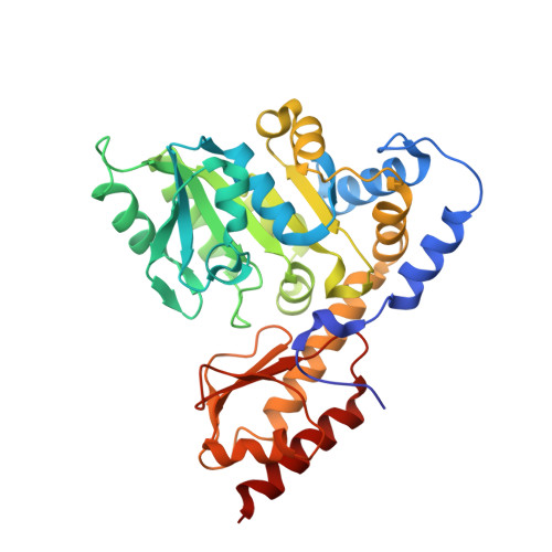 | |
UniProt | |||||
Find proteins for A0A166JNM8 (Pseudomonas putida) Explore A0A166JNM8 Go to UniProtKB: A0A166JNM8 | |||||
Entity Groups | |||||
| Sequence Clusters | 30% Identity50% Identity70% Identity90% Identity95% Identity100% Identity | ||||
| UniProt Group | A0A166JNM8 | ||||
Sequence AnnotationsExpand | |||||
| |||||
| Ligands 2 Unique | |||||
|---|---|---|---|---|---|
| ID | Chains | Name / Formula / InChI Key | 2D Diagram | 3D Interactions | |
| PLR Query on PLR | E [auth A], G [auth B], I [auth C], K [auth D] | (5-HYDROXY-4,6-DIMETHYLPYRIDIN-3-YL)METHYL DIHYDROGEN PHOSPHATE C8 H12 N O5 P RBCOYOYDYNXAFA-UHFFFAOYSA-N |  | ||
| GOL Query on GOL | F [auth A], H [auth B], J [auth C], L [auth D] | GLYCEROL C3 H8 O3 PEDCQBHIVMGVHV-UHFFFAOYSA-N |  | ||
| Length ( Å ) | Angle ( ˚ ) |
|---|---|
| a = 198.167 | α = 90 |
| b = 186.96 | β = 98.92 |
| c = 53.269 | γ = 90 |
| Software Name | Purpose |
|---|---|
| PHENIX | refinement |
| Coot | model building |
| XDS | data reduction |
| PHENIX | phasing |
RCSB PDB (citation) is hosted by
RCSB PDB is a member of the

