5GWF

FraC with GlcNAc(6S) bound


Experimental Data Snapshot

  • Method: X-RAY DIFFRACTION
  • Resolution: 1.55 Å
  • R-Value Free: 0.163 
  • R-Value Work: 0.118 
  • R-Value Observed: 0.119 

wwPDB Validation   3D Report Full Report


Ligand Structure Quality Assessment 


This is version 2.1 of the entry. See complete history


Literature

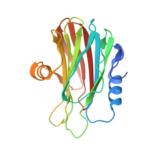
Haemolytic actinoporins interact with carbohydrates using their lipid-binding module

Tanaka, K.Caaveiro, J.M.M.Morante, K.Tsumoto, K.

(2017) Philos Trans R Soc Lond B Biol Sci 372

  • DOI: https://doi.org/10.1098/rstb.2016.0216
  • Primary Citation of Related Structures:  
    5GWF

  • PubMed Abstract: 

    Pore-forming toxins (PFTs) are proteins endowed with metamorphic properties that enable them to stably fold in water solutions as well as in cellular membranes. PFTs produce lytic pores on the plasma membranes of target cells conducive to lesions, playing key roles in the defensive and offensive molecular systems of living organisms. Actinoporins are a family of potent haemolytic toxins produced by sea anemones vigorously studied as a paradigm of α-helical PFTs, in the context of lipid-protein interactions, and in connection with nanopore technologies. We have recently reported that fragaceatoxin C (FraC), an actinoporin, engages biological membranes with a large adhesive motif allowing the simultaneous attachment of up to four lipid molecules prior to pore formation. Since actinoporins also interact with carbohydrates, we sought to understand the molecular and energetic basis of glycan recognition by FraC. By employing structural and biophysical methodologies, we show that FraC engages glycans with low affinity using its lipid-binding module. Contrary to other PFTs requiring separate domains for glycan and lipid recognition, the small single-domain actinoporins economize resources by achieving dual recognition with a single binding module. This mechanism could enhance the recruitment of actinoporins to the surface of target tissues in their marine environment.This article is part of the themed issue 'Membrane pores: from structure and assembly, to medicine and technology'.


  • Organizational Affiliation

    Department of Chemistry and Biotechnology, The University of Tokyo, Bunkyo-ku, Tokyo 113-8656, Japan.


Macromolecules
Find similar proteins by:  (by identity cutoff)  |  3D Structure
Entity ID: 1
MoleculeChains Sequence LengthOrganismDetailsImage
DELTA-actitoxin-Afr1a
A, B, C, D
180Actinia fragaceaMutation(s): 0 
UniProt
Find proteins for B9W5G6 (Actinia fragacea)
Explore B9W5G6 
Go to UniProtKB:  B9W5G6
Entity Groups  
Sequence Clusters30% Identity50% Identity70% Identity90% Identity95% Identity100% Identity
UniProt GroupB9W5G6
Sequence Annotations
Expand
  • Reference Sequence
Small Molecules
Ligands 3 Unique
IDChains Name / Formula / InChI Key2D Diagram3D Interactions
NGY
Query on NGY

Download Ideal Coordinates CCD File 
F [auth B],
R [auth D]
2-acetamido-2-deoxy-6-O-sulfo-alpha-D-glucopyranose
C8 H15 N O9 S
WJFVEEAIYIOATH-PVFLNQBWSA-N
GOL
Query on GOL

Download Ideal Coordinates CCD File 
M [auth B],
Q [auth C]
GLYCEROL
C3 H8 O3
PEDCQBHIVMGVHV-UHFFFAOYSA-N
CL
Query on CL

Download Ideal Coordinates CCD File 
E [auth A]
G [auth B]
H [auth B]
I [auth B]
J [auth B]
E [auth A],
G [auth B],
H [auth B],
I [auth B],
J [auth B],
K [auth B],
L [auth B],
N [auth C],
O [auth C],
P [auth C],
S [auth D],
T [auth D],
U [auth D]
CHLORIDE ION
Cl
VEXZGXHMUGYJMC-UHFFFAOYSA-M
Experimental Data & Validation

Experimental Data

  • Method: X-RAY DIFFRACTION
  • Resolution: 1.55 Å
  • R-Value Free: 0.163 
  • R-Value Work: 0.118 
  • R-Value Observed: 0.119 
  • Space Group: P 1 21 1
Unit Cell:
Length ( Å )Angle ( ˚ )
a = 77.16α = 90
b = 44.4β = 92.76
c = 114.36γ = 90
Software Package:
Software NamePurpose
REFMACrefinement
MOSFLMdata reduction
SCALAdata scaling
PHASERphasing

Structure Validation

View Full Validation Report


Ligand Structure Quality Assessment 


Entry History & Funding Information

Deposition Data


Funding OrganizationLocationGrant Number
JSPSJapan25249115
JSPSJapan15K06962

Revision History  (Full details and data files)

  • Version 1.0: 2017-06-21
    Type: Initial release
  • Version 1.1: 2017-12-06
    Changes: Database references
  • Version 2.0: 2020-07-29
    Type: Remediation
    Reason: Carbohydrate remediation
    Changes: Atomic model, Data collection, Derived calculations, Structure summary
  • Version 2.1: 2023-11-08
    Changes: Data collection, Database references, Refinement description, Structure summary