5E85

isolated SBD of BiP


Experimental Data Snapshot

  • Method: X-RAY DIFFRACTION
  • Resolution: 2.57 Å
  • R-Value Free: 0.264 
  • R-Value Work: 0.219 
  • R-Value Observed: 0.222 

wwPDB Validation   3D Report Full Report


This is version 1.3 of the entry. See complete history


Literature

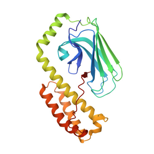
Close and Allosteric Opening of the Polypeptide-Binding Site in a Human Hsp70 Chaperone BiP.

Yang, J.Nune, M.Zong, Y.Zhou, L.Liu, Q.

(2015) Structure 23: 2191-2203

  • DOI: https://doi.org/10.1016/j.str.2015.10.012
  • Primary Citation of Related Structures:  
    5E84, 5E85, 5E86

  • PubMed Abstract: 

    Binding immunoglobulin protein (BiP), an essential and ubiquitous Hsp70 chaperone in the ER, plays a key role in protein folding and quality control. BiP contains two functional domains: a nucleotide-binding domain (NBD) and a substrate-binding domain (SBD). NBD binds and hydrolyzes ATP; the substrates for SBD are extended polypeptides. ATP binding allosterically accelerates polypeptide binding and release. Although crucial to the chaperone activity, the molecular mechanisms of polypeptide binding and allosteric coupling of BiP are poorly understood. Here, we present crystal structures of an intact human BiP in the ATP-bound state, the first intact eukaryotic Hsp70 structure, and isolated BiP-SBD with a peptide substrate bound representing the ADP-bound state. These structures and our biochemical analysis demonstrate that BiP has a unique NBD-SBD interface that is highly conserved only in eukaryotic Hsp70s found in the cytosol and ER to fortify its ATP-bound state and promote the opening of its polypeptide-binding pocket.


  • Organizational Affiliation

    Department of Physiology and Biophysics, Virginia Commonwealth University, Richmond, VA 23298, USA.


Macromolecules
Find similar proteins by:  (by identity cutoff)  |  3D Structure
Entity ID: 1
MoleculeChains Sequence LengthOrganismDetailsImage
78 kDa glucose-regulated protein236Homo sapiensMutation(s): 0 
Gene Names: HSPA5GRP78
UniProt & NIH Common Fund Data Resources
Find proteins for P11021 (Homo sapiens)
Explore P11021 
Go to UniProtKB:  P11021
PHAROS:  P11021
GTEx:  ENSG00000044574 
Entity Groups  
Sequence Clusters30% Identity50% Identity70% Identity90% Identity95% Identity100% Identity
UniProt GroupP11021
Sequence Annotations
Expand
  • Reference Sequence
Small Molecules
Ligands 1 Unique
IDChains Name / Formula / InChI Key2D Diagram3D Interactions
SO4
Query on SO4

Download Ideal Coordinates CCD File 
B [auth A]SULFATE ION
O4 S
QAOWNCQODCNURD-UHFFFAOYSA-L
Experimental Data & Validation

Experimental Data

  • Method: X-RAY DIFFRACTION
  • Resolution: 2.57 Å
  • R-Value Free: 0.264 
  • R-Value Work: 0.219 
  • R-Value Observed: 0.222 
  • Space Group: C 2 2 21
Unit Cell:
Length ( Å )Angle ( ˚ )
a = 36.378α = 90
b = 91.661β = 90
c = 141.797γ = 90
Software Package:
Software NamePurpose
PHENIXrefinement
HKL-2000data reduction
HKL-2000data scaling
PHASERphasing

Structure Validation

View Full Validation Report


Entry History & Funding Information

Deposition Data


Funding OrganizationLocationGrant Number
National Institutes of Health/National Institute of General Medical Sciences (NIH/NIGMS)United States1R01GM098592, 1RO1GM109193

Revision History  (Full details and data files)

  • Version 1.0: 2015-12-30
    Type: Initial release
  • Version 1.1: 2017-09-20
    Changes: Author supporting evidence, Database references, Derived calculations
  • Version 1.2: 2019-12-25
    Changes: Author supporting evidence
  • Version 1.3: 2024-03-06
    Changes: Data collection, Database references