4Z45

Structure of OBP3 from the currant-lettuce aphid Nasonovia ribisnigri


Experimental Data Snapshot

  • Method: X-RAY DIFFRACTION
  • Resolution: 2.02 Å
  • R-Value Free: 0.207 
  • R-Value Work: 0.164 
  • R-Value Observed: 0.166 

wwPDB Validation   3D Report Full Report


This is version 1.4 of the entry. See complete history


Literature

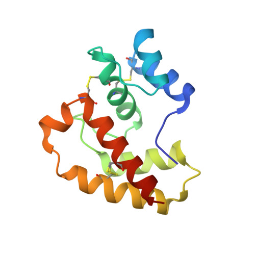
Crystal Structures and Binding Dynamics of Odorant-Binding Protein 3 from two aphid species Megoura viciae and Nasonovia ribisnigri.

Northey, T.Venthur, H.De Biasio, F.Chauviac, F.X.Cole, A.Ribeiro, K.A.Grossi, G.Falabella, P.Field, L.M.Keep, N.H.Zhou, J.J.

(2016) Sci Rep 6: 24739-24739

  • DOI: https://doi.org/10.1038/srep24739
  • Primary Citation of Related Structures:  
    4Z39, 4Z45

  • PubMed Abstract: 

    Aphids use chemical cues to locate hosts and find mates. The vetch aphid Megoura viciae feeds exclusively on the Fabaceae, whereas the currant-lettuce aphid Nasonovia ribisnigri alternates hosts between the Grossulariaceae and Asteraceae. Both species use alarm pheromones to warn of dangers. For N. ribisnigri this pheromone is a single component (E)-β-farnesene but M. viciae uses a mixture of (E)-β-farnesene, (-)-α-pinene, β-pinene, and limonene. Odorant-binding proteins (OBP) are believed to capture and transport such semiochemicals to their receptors. Here, we report the first aphid OBP crystal structures and examine their molecular interactions with the alarm pheromone components. Our study reveals some unique structural features: 1) the lack of an internal ligand binding site; 2) a striking groove in the surface of the proteins as a putative binding site; 3) the N-terminus rather than the C-terminus occupies the site closing off the conventional OBP pocket. The results from fluorescent binding assays, molecular docking and dynamics demonstrate that OBP3 from M. viciae can bind to all four alarm pheromone components and the differential ligand binding between these very similar OBP3s from the two aphid species is determined mainly by the direct π-π interactions between ligands and the aromatic residues of OBP3s in the binding pocket.


  • Organizational Affiliation

    Crystallography, Institute for Structural and Molecular Biology, Department of Biological Sciences, Birkbeck, University of London, London WC1E 7HX, UK.


Macromolecules
Find similar proteins by:  (by identity cutoff)  |  3D Structure
Entity ID: 1
MoleculeChains Sequence LengthOrganismDetailsImage
Odorant-binding protein NribOBP3A,
B,
C [auth D]
121Nasonovia ribisnigriMutation(s): 0 
Gene Names: obp3
UniProt
Find proteins for A0A0M4AUH6 (Nasonovia ribisnigri)
Explore A0A0M4AUH6 
Go to UniProtKB:  A0A0M4AUH6
Entity Groups  
Sequence Clusters30% Identity50% Identity70% Identity90% Identity95% Identity100% Identity
UniProt GroupA0A0M4AUH6
Sequence Annotations
Expand
  • Reference Sequence
Experimental Data & Validation

Experimental Data

  • Method: X-RAY DIFFRACTION
  • Resolution: 2.02 Å
  • R-Value Free: 0.207 
  • R-Value Work: 0.164 
  • R-Value Observed: 0.166 
  • Space Group: P 31 2 1
Unit Cell:
Length ( Å )Angle ( ˚ )
a = 87.25α = 90
b = 87.25β = 90
c = 95.08γ = 120
Software Package:
Software NamePurpose
REFMACrefinement
XDSdata reduction
XSCALEdata scaling
PHASERphasing

Structure Validation

View Full Validation Report


Entry History & Funding Information

Deposition Data


Funding OrganizationLocationGrant Number
Biotechnology and Biological Sciences Research CouncilUnited KingdomBB/L001683/1
Biotechnology and Biological Sciences Research CouncilUnited KingdomBB/I024941/1

Revision History  (Full details and data files)

  • Version 1.0: 2016-04-13
    Type: Initial release
  • Version 1.1: 2016-04-20
    Changes: Database references
  • Version 1.2: 2016-07-27
    Changes: Database references
  • Version 1.3: 2017-08-30
    Changes: Advisory, Author supporting evidence, Derived calculations
  • Version 1.4: 2019-05-08
    Changes: Data collection, Derived calculations