4XWM

Complex structure of catalytic domain of Clostridium Cellulovorans Exgs and Cellobiose


Experimental Data Snapshot

  • Method: X-RAY DIFFRACTION
  • Resolution: 1.70 Å
  • R-Value Free: 0.163 
  • R-Value Work: 0.146 
  • R-Value Observed: 0.146 

wwPDB Validation   3D Report Full Report


This is version 2.1 of the entry. See complete history


Literature

Structures of exoglucanase from Clostridium cellulovorans: cellotetraose binding and cleavage

Tsai, L.-C.Amiraslanov, I.Chen, H.R.Chen, Y.W.Lee, H.L.Liang, P.H.Liaw, Y.-C.

(2015) Acta Crystallogr F Struct Biol Commun 71: 1264-1272

  • DOI: https://doi.org/10.1107/S2053230X15015915
  • Primary Citation of Related Structures:  
    4XWL, 4XWM, 4XWN

  • PubMed Abstract: 

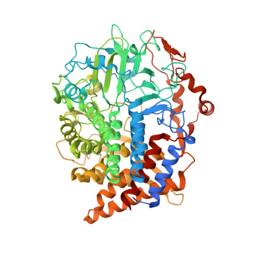
    Exoglucanase/cellobiohydrolase (EC 3.2.1.176) hydrolyzes a β-1,4-glycosidic bond from the reducing end of cellulose and releases cellobiose as the major product. Three complex crystal structures of the glycosyl hydrolase 48 (GH48) cellobiohydrolase S (ExgS) from Clostridium cellulovorans with cellobiose, cellotetraose and triethylene glycol molecules were solved. The product cellobiose occupies subsites +1 and +2 in the open active-site cleft of the enzyme-cellotetraose complex structure, indicating an enzymatic hydrolysis function. Moreover, three triethylene glycol molecules and one pentaethylene glycol molecule are located at active-site subsites -2 to -6 in the structure of the ExgS-triethylene glycol complex shown here. Modelling of glucose into subsite -1 in the active site of the ExgS-cellobiose structure revealed that Glu50 acts as a proton donor and Asp222 plays a nucleophilic role.


  • Organizational Affiliation

    Molecular Science and Engineering, National Taipei University of Technology, 1, Section 3, Chung-Hsiao E. Road, Taipei 10608, Taiwan.


Macromolecules
Find similar proteins by:  (by identity cutoff)  |  3D Structure
Entity ID: 1
MoleculeChains Sequence LengthOrganismDetailsImage
Exoglucanase S681Clostridium cellulovoransMutation(s): 0 
Gene Names: exgS
EC: 3.2.1.91
UniProt
Find proteins for O65986 (Clostridium cellulovorans)
Explore O65986 
Go to UniProtKB:  O65986
Entity Groups  
Sequence Clusters30% Identity50% Identity70% Identity90% Identity95% Identity100% Identity
UniProt GroupO65986
Sequence Annotations
Expand
  • Reference Sequence
Oligosaccharides

Help

Entity ID: 2
MoleculeChains Length2D Diagram Glycosylation3D Interactions
beta-D-glucopyranose-(1-4)-alpha-D-glucopyranose
B
2N/A
Glycosylation Resources
GlyTouCan:  G86802XL
GlyCosmos:  G86802XL
Entity ID: 3
MoleculeChains Length2D Diagram Glycosylation3D Interactions
beta-D-glucopyranose-(1-4)-beta-D-glucopyranose
C, D
2N/A
Glycosylation Resources
GlyTouCan:  G84824ZO
GlyCosmos:  G84824ZO
Experimental Data & Validation

Experimental Data

  • Method: X-RAY DIFFRACTION
  • Resolution: 1.70 Å
  • R-Value Free: 0.163 
  • R-Value Work: 0.146 
  • R-Value Observed: 0.146 
  • Space Group: P 41 21 2
Unit Cell:
Length ( Å )Angle ( ˚ )
a = 109.14α = 90
b = 109.14β = 90
c = 182.145γ = 90
Software Package:
Software NamePurpose
SCALEPACKdata scaling
PHASERphasing
RESOLVEphasing
PHENIXrefinement
PDB_EXTRACTdata extraction

Structure Validation

View Full Validation Report


Entry History 

Deposition Data

  • Released Date: 2015-10-28 
  • Deposition Author(s): Liaw, Y.-C.
  • This entry supersedes: 4KKF

Revision History  (Full details and data files)

  • Version 1.0: 2015-10-28
    Type: Initial release
  • Version 2.0: 2020-07-29
    Type: Remediation
    Reason: Carbohydrate remediation
    Changes: Atomic model, Data collection, Derived calculations, Non-polymer description, Structure summary
  • Version 2.1: 2023-11-08
    Changes: Data collection, Database references, Refinement description, Structure summary