4X8Y

Crystal structure of human PGRMC1 cytochrome b5-like domain


Experimental Data Snapshot

  • Method: X-RAY DIFFRACTION
  • Resolution: 1.95 Å
  • R-Value Free: 0.212 
  • R-Value Work: 0.183 
  • R-Value Observed: 0.185 

wwPDB Validation   3D Report Full Report


Ligand Structure Quality Assessment 


This is version 1.2 of the entry. See complete history


Literature

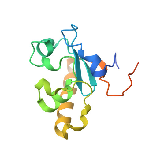
Haem-dependent dimerization of PGRMC1/Sigma-2 receptor facilitates cancer proliferation and chemoresistance

Kabe, Y.Nakane, T.Koike, I.Yamamoto, T.Sugiura, Y.Harada, E.Sugase, K.Shimamura, T.Ohmura, M.Muraoka, K.Yamamoto, A.Uchida, T.Iwata, S.Yamaguchi, Y.Krayukhina, E.Noda, M.Handa, H.Ishimori, K.Uchiyama, S.Kobayashi, T.Suematsu, M.

(2016) Nat Commun 7: 11030-11030

  • DOI: https://doi.org/10.1038/ncomms11030
  • Primary Citation of Related Structures:  
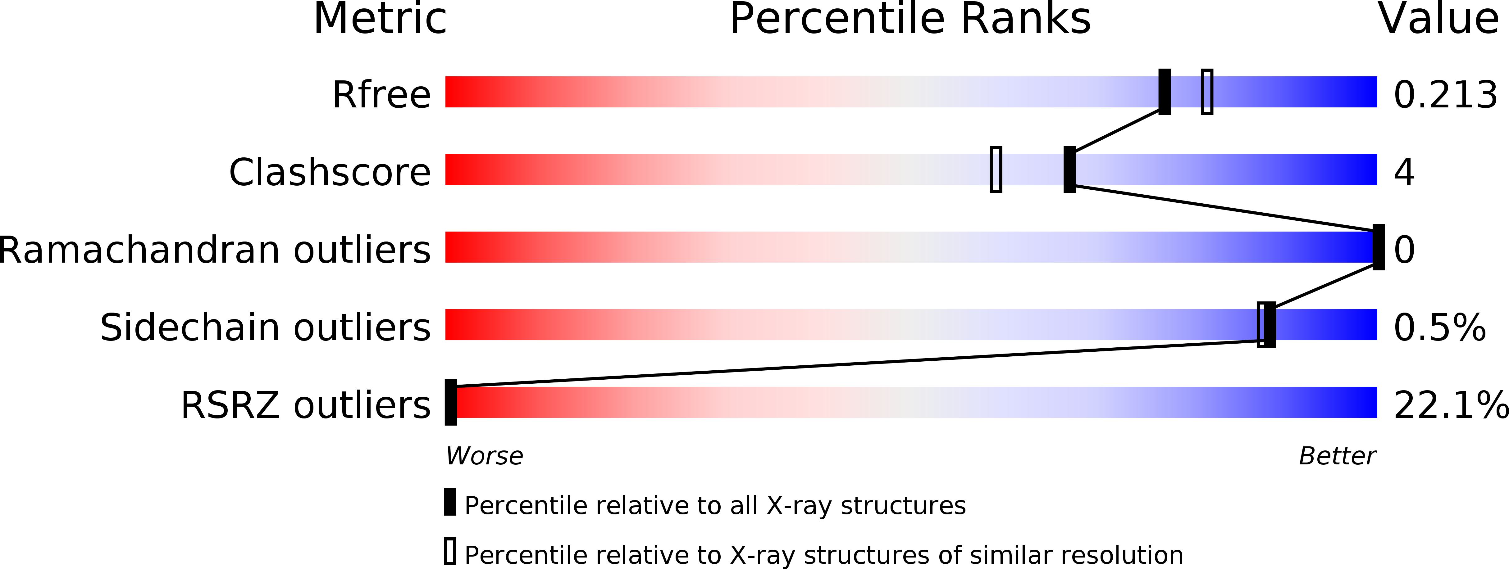
    4X8Y

  • PubMed Abstract: 

    Progesterone-receptor membrane component 1 (PGRMC1/Sigma-2 receptor) is a haem-containing protein that interacts with epidermal growth factor receptor (EGFR) and cytochromes P450 to regulate cancer proliferation and chemoresistance; its structural basis remains unknown. Here crystallographic analyses of the PGRMC1 cytosolic domain at 1.95 Å resolution reveal that it forms a stable dimer through stacking interactions of two protruding haem molecules. The haem iron is five-coordinated by Tyr113, and the open surface of the haem mediates dimerization. Carbon monoxide (CO) interferes with PGRMC1 dimerization by binding to the sixth coordination site of the haem. Haem-mediated PGRMC1 dimerization is required for interactions with EGFR and cytochromes P450, cancer proliferation and chemoresistance against anti-cancer drugs; these events are attenuated by either CO or haem deprivation in cancer cells. This study demonstrates protein dimerization via haem-haem stacking, which has not been seen in eukaryotes, and provides insights into its functional significance in cancer.


  • Organizational Affiliation

    Department of Biochemistry, Keio University School of Medicine, and Japan Science and Technology Agency (JST), Core Research for Evolutional Science and Technology (CREST), Tokyo 160-8582, Japan.


Macromolecules
Find similar proteins by:  (by identity cutoff)  |  3D Structure
Entity ID: 1
MoleculeChains Sequence LengthOrganismDetailsImage
Membrane-associated progesterone receptor component 1
A, B
132Homo sapiensMutation(s): 0 
Gene Names: PGRMC1HPR6.6PGRMC
UniProt & NIH Common Fund Data Resources
Find proteins for O00264 (Homo sapiens)
Explore O00264 
Go to UniProtKB:  O00264
PHAROS:  O00264
GTEx:  ENSG00000101856 
Entity Groups  
Sequence Clusters30% Identity50% Identity70% Identity90% Identity95% Identity100% Identity
UniProt GroupO00264
Sequence Annotations
Expand
  • Reference Sequence
Small Molecules
Ligands 1 Unique
IDChains Name / Formula / InChI Key2D Diagram3D Interactions
HEM
Query on HEM

Download Ideal Coordinates CCD File 
C [auth A],
D [auth B]
PROTOPORPHYRIN IX CONTAINING FE
C34 H32 Fe N4 O4
KABFMIBPWCXCRK-RGGAHWMASA-L
Experimental Data & Validation

Experimental Data

  • Method: X-RAY DIFFRACTION
  • Resolution: 1.95 Å
  • R-Value Free: 0.212 
  • R-Value Work: 0.183 
  • R-Value Observed: 0.185 
  • Space Group: I 41 2 2
Unit Cell:
Length ( Å )Angle ( ˚ )
a = 167.23α = 90
b = 167.23β = 90
c = 63.46γ = 90
Software Package:
Software NamePurpose
PHENIXmodel building
PHENIXrefinement
XDSdata reduction
XSCALEdata scaling
PHENIXphasing
BSSdata collection

Structure Validation

View Full Validation Report


Ligand Structure Quality Assessment 


Entry History & Funding Information

Deposition Data


Funding OrganizationLocationGrant Number
Japan Science and Technology AgencyJapanExploratory Research for Advanced Technology (ERATO) Suematsu Gas Biology Project

Revision History  (Full details and data files)

  • Version 1.0: 2016-03-23
    Type: Initial release
  • Version 1.1: 2016-03-30
    Changes: Database references
  • Version 1.2: 2020-02-05
    Changes: Data collection, Derived calculations