4X1T

The crystal structure of Arabidopsis thaliana galactolipid synthase MGD1 in complex with UDP


Experimental Data Snapshot

  • Method: X-RAY DIFFRACTION
  • Resolution: 2.25 Å
  • R-Value Free: 0.216 
  • R-Value Work: 0.199 
  • R-Value Observed: 0.199 

wwPDB Validation   3D Report Full Report


Ligand Structure Quality Assessment 


This is version 2.1 of the entry. See complete history


Literature

Structural insights and membrane binding properties of MGD1, the major galactolipid synthase in plants.

Rocha, J.Sarkis, J.Thomas, A.Pitou, L.Radzimanowski, J.Audry, M.Chazalet, V.de Sanctis, D.Palcic, M.M.Block, M.A.Girard-Egrot, A.Marechal, E.Breton, C.

(2016) Plant J 85: 622-633

  • DOI: https://doi.org/10.1111/tpj.13129
  • Primary Citation of Related Structures:  
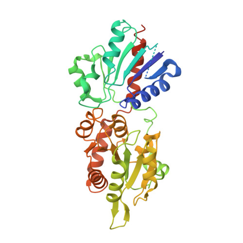
    4WYI, 4X1T

  • PubMed Abstract: 

    Monogalactosyldiacylglycerol (MGDG) and digalactosyldiacylglycerol (DGDG) are the major lipid components of photosynthetic membranes, and hence the most abundant lipids in the biosphere. They are essential for assembly and function of the photosynthetic apparatus. In Arabidopsis, the first step of galactolipid synthesis is catalyzed by MGDG synthase 1 (MGD1), which transfers a galactosyl residue from UDP-galactose to diacylglycerol (DAG). MGD1 is a monotopic protein that is embedded in the inner envelope membrane of chloroplasts. Once produced, MGDG is transferred to the outer envelope membrane, where DGDG synthesis occurs, and to thylakoids. Here we present two crystal structures of MGD1: one unliganded and one complexed with UDP. MGD1 has a long and flexible region (approximately 50 amino acids) that is required for DAG binding. The structures reveal critical features of the MGD1 catalytic mechanism and its membrane binding mode, tested on biomimetic Langmuir monolayers, giving insights into chloroplast membrane biogenesis. The structural plasticity of MGD1, ensuring very rapid capture and utilization of DAG, and its interaction with anionic lipids, possibly driving the construction of lipoproteic clusters, are consistent with the role of this enzyme, not only in expansion of the inner envelope membrane, but also in supplying MGDG to the outer envelope and nascent thylakoid membranes.


  • Organizational Affiliation

    University of Grenoble Alpes, 38400, Grenoble, France.


Macromolecules
Find similar proteins by:  (by identity cutoff)  |  3D Structure
Entity ID: 1
MoleculeChains Sequence LengthOrganismDetailsImage
Monogalactosyldiacylglycerol synthase 1, chloroplastic408Arabidopsis thalianaMutation(s): 0 
Gene Names: MGD1MGDAAt4g31780F28M20.30
EC: 2.4.1.46
UniProt
Find proteins for O81770 (Arabidopsis thaliana)
Explore O81770 
Go to UniProtKB:  O81770
Entity Groups  
Sequence Clusters30% Identity50% Identity70% Identity90% Identity95% Identity100% Identity
UniProt GroupO81770
Sequence Annotations
Expand
  • Reference Sequence
Small Molecules
Ligands 2 Unique
IDChains Name / Formula / InChI Key2D Diagram3D Interactions
UDP
Query on UDP

Download Ideal Coordinates CCD File 
B [auth A]URIDINE-5'-DIPHOSPHATE
C9 H14 N2 O12 P2
XCCTYIAWTASOJW-XVFCMESISA-N
EDO
Query on EDO

Download Ideal Coordinates CCD File 
C [auth A]
D [auth A]
E [auth A]
F [auth A]
G [auth A]
C [auth A],
D [auth A],
E [auth A],
F [auth A],
G [auth A],
H [auth A],
I [auth A],
J [auth A],
K [auth A]
1,2-ETHANEDIOL
C2 H6 O2
LYCAIKOWRPUZTN-UHFFFAOYSA-N
Experimental Data & Validation

Experimental Data

  • Method: X-RAY DIFFRACTION
  • Resolution: 2.25 Å
  • R-Value Free: 0.216 
  • R-Value Work: 0.199 
  • R-Value Observed: 0.199 
  • Space Group: P 63
Unit Cell:
Length ( Å )Angle ( ˚ )
a = 139.25α = 90
b = 139.25β = 90
c = 38.27γ = 120
Software Package:
Software NamePurpose
PHENIXrefinement
XDSdata reduction
XSCALEdata scaling
PHASERphasing

Structure Validation

View Full Validation Report


Ligand Structure Quality Assessment 


Entry History & Funding Information

Deposition Data


Funding OrganizationLocationGrant Number
French National Research AgencyFranceANR-10-BLANC-1524

Revision History  (Full details and data files)

  • Version 1.0: 2016-02-10
    Type: Initial release
  • Version 1.1: 2016-10-05
    Changes: Database references
  • Version 2.0: 2017-08-30
    Changes: Advisory, Atomic model, Author supporting evidence, Data collection, Derived calculations
  • Version 2.1: 2024-01-10
    Changes: Data collection, Database references, Refinement description