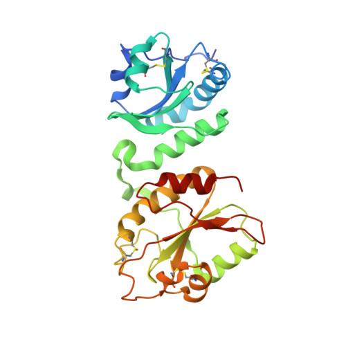
The eps1p protein disulfide isomerase conserves classic thioredoxin superfamily amino Acid motifs but not their functional geometries.
Biran, S., Gat, Y., Fass, D.(2014) PLoS One 9: e113431-e113431
- PubMed: 25437863
- DOI: https://doi.org/10.1371/journal.pone.0113431
- Primary Citation of Related Structures:
4TVE, 4TW5 - PubMed Abstract:
The widespread thioredoxin superfamily enzymes typically share the following features: a characteristic α-β fold, the presence of a Cys-X-X-Cys (or Cys-X-X-Ser) redox-active motif, and a proline in the cis configuration abutting the redox-active site in the tertiary structure. The Cys-X-X-Cys motif is at the solvent-exposed amino terminus of an α-helix, allowing the first cysteine to engage in nucleophilic attack on substrates, or substrates to attack the Cys-X-X-Cys disulfide, depending on whether the enzyme functions to reduce, isomerize, or oxidize its targets. We report here the X-ray crystal structure of an enzyme that breaks many of our assumptions regarding the sequence-structure relationship of thioredoxin superfamily proteins. The yeast Protein Disulfide Isomerase family member Eps1p has Cys-X-X-Cys motifs and proline residues at the appropriate primary structural positions in its first two predicted thioredoxin-fold domains. However, crystal structures show that the Cys-X-X-Cys of the second domain is buried and that the adjacent proline is in the trans, rather than the cis isomer. In these configurations, neither the "active-site" disulfide nor the backbone carbonyl preceding the proline is available to interact with substrate. The Eps1p structures thus expand the documented diversity of the PDI oxidoreductase family and demonstrate that conserved sequence motifs in common folds do not guarantee structural or functional conservation.
- Department of Structural Biology, Weizmann Institute of Science, Rehovot, Israel.
Organizational Affiliation: