4TN7

Crystal structure of mouse KDM2A-H3K36ME-NO complex

  • Classification: OXIDOREDUCTASE
  • Organism(s): Mus musculus, Homo sapiens
  • Expression System: Escherichia coli BL21(DE3)
  • Mutation(s): No 

  • Deposited: 2014-06-03 Released: 2015-05-13 
  • Deposition Author(s): Cheng, Z.
  • Funding Organization(s): Lymphoma Society and the STARR foundation, National Institutes of Health/National Institute of General Medical Sciences (NIH/NIGMS)

Experimental Data Snapshot

  • Method: X-RAY DIFFRACTION
  • Resolution: 2.20 Å
  • R-Value Free: 0.279 
  • R-Value Work: 0.229 
  • R-Value Observed: 0.232 

wwPDB Validation   3D Report Full Report


This is version 1.3 of the entry. See complete history


Literature

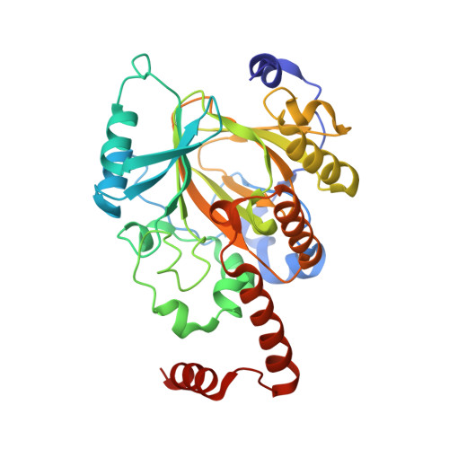
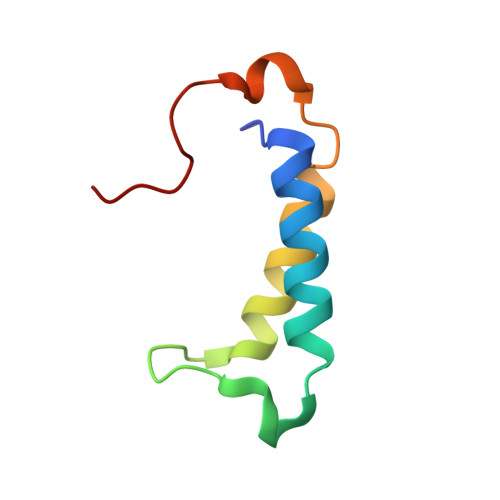
A molecular threading mechanism underlies Jumonji lysine demethylase KDM2A regulation of methylated H3K36.

Cheng, Z.Cheung, P.Kuo, A.J.Yukl, E.T.Wilmot, C.M.Gozani, O.Patel, D.J.

(2014) Genes Dev 28: 1758-1771

  • DOI: https://doi.org/10.1101/gad.246561.114
  • Primary Citation of Related Structures:  
    4QWN, 4QX7, 4QX8, 4QXB, 4QXC, 4QXH, 4TN7

  • PubMed Abstract: 

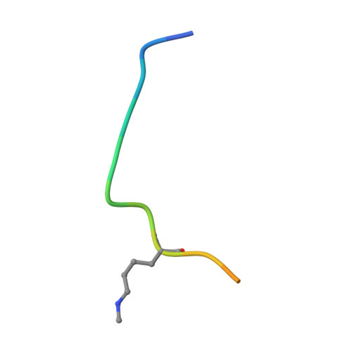
    The dynamic reversible methylation of lysine residues on histone proteins is central to chromatin biology. Key components are demethylase enzymes, which remove methyl moieties from lysine residues. KDM2A, a member of the Jumonji C domain-containing histone lysine demethylase family, specifically targets lower methylation states of H3K36. Here, structural studies reveal that H3K36 specificity for KDM2A is mediated by the U-shaped threading of the H3K36 peptide through a catalytic groove within KDM2A. The side chain of methylated K36 inserts into the catalytic pocket occupied by Ni(2+) and cofactor, where it is positioned and oriented for demethylation. Key residues contributing to K36me specificity on histone H3 are G33 and G34 (positioned within a narrow channel), P38 (a turn residue), and Y41 (inserts into its own pocket). Given that KDM2A was found to also bind the H3K36me3 peptide, we postulate that steric constraints could prevent α-ketoglutarate from undergoing an "off-line"-to-"in-line" transition necessary for the demethylation reaction. Furthermore, structure-guided substitutions of residues in the KDM2A catalytic pocket abrogate KDM2A-mediated functions important for suppression of cancer cell phenotypes. Together, our results deduce insights into the molecular basis underlying KDM2A regulation of the biologically important methylated H3K36 mark.


  • Organizational Affiliation

    Structural Biology Program, Memorial Sloan-Kettering Cancer Center, New York, New York 10065, USA;


Macromolecules
Find similar proteins by:  (by identity cutoff)  |  3D Structure
Entity ID: 1
MoleculeChains Sequence LengthOrganismDetailsImage
Lysine-specific demethylase 2A
A, C
329Mus musculusMutation(s): 0 
Gene Names: Kdm2aFbxl11Jhdm1aKiaa1004
EC: 1.14.11.27
UniProt
Find proteins for P59997 (Mus musculus)
Explore P59997 
Go to UniProtKB:  P59997
Entity Groups  
Sequence Clusters30% Identity50% Identity70% Identity90% Identity95% Identity100% Identity
UniProt GroupP59997
Sequence Annotations
Expand
  • Reference Sequence
Find similar proteins by:  (by identity cutoff)  |  3D Structure
Entity ID: 2
MoleculeChains Sequence LengthOrganismDetailsImage
Lysine-specific demethylase 2A
B, D
68Mus musculusMutation(s): 0 
Gene Names: Kdm2aFbxl11Jhdm1aKiaa1004
EC: 1.14.11.27
UniProt
Find proteins for P59997 (Mus musculus)
Explore P59997 
Go to UniProtKB:  P59997
Entity Groups  
Sequence Clusters30% Identity50% Identity70% Identity90% Identity95% Identity100% Identity
UniProt GroupP59997
Sequence Annotations
Expand
  • Reference Sequence

Find similar proteins by:  Sequence   |   3D Structure  

Entity ID: 3
MoleculeChains Sequence LengthOrganismDetailsImage
Peptide
E, F
15Homo sapiensMutation(s): 0 
UniProt & NIH Common Fund Data Resources
Find proteins for P68431 (Homo sapiens)
Explore P68431 
Go to UniProtKB:  P68431
PHAROS:  P68431
Entity Groups  
Sequence Clusters30% Identity50% Identity70% Identity90% Identity95% Identity100% Identity
UniProt GroupP68431
Sequence Annotations
Expand
  • Reference Sequence
Experimental Data & Validation

Experimental Data

  • Method: X-RAY DIFFRACTION
  • Resolution: 2.20 Å
  • R-Value Free: 0.279 
  • R-Value Work: 0.229 
  • R-Value Observed: 0.232 
  • Space Group: P 1 21 1
Unit Cell:
Length ( Å )Angle ( ˚ )
a = 55.063α = 90
b = 157.928β = 90
c = 48.706γ = 90
Software Package:
Software NamePurpose
PHENIXrefinement

Structure Validation

View Full Validation Report


Entry History & Funding Information

Deposition Data

  • Released Date: 2015-05-13 
  • Deposition Author(s): Cheng, Z.

Funding OrganizationLocationGrant Number
Lymphoma Society and the STARR foundationUnited States--
National Institutes of Health/National Institute of General Medical Sciences (NIH/NIGMS)United StatesR01 GM079641
National Institutes of Health/National Institute of General Medical Sciences (NIH/NIGMS)United StatesR01 GM066569
National Institutes of Health/National Institute of General Medical Sciences (NIH/NIGMS)United StatesF32 GM097779

Revision History  (Full details and data files)

  • Version 1.0: 2015-05-13
    Type: Initial release
  • Version 1.1: 2017-09-20
    Changes: Advisory, Author supporting evidence, Database references, Derived calculations, Source and taxonomy, Structure summary
  • Version 1.2: 2019-12-25
    Changes: Author supporting evidence
  • Version 1.3: 2023-09-27
    Changes: Advisory, Data collection, Database references, Derived calculations, Refinement description