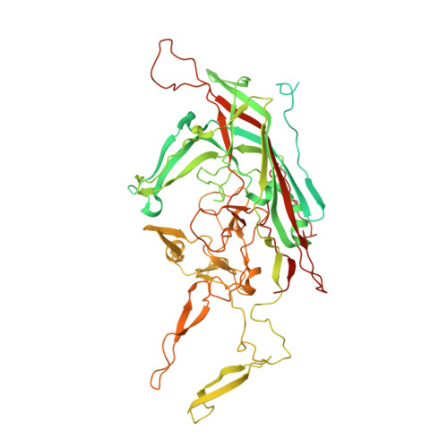
Structure of neurotropic adeno-associated virus AAVrh.8.
Halder, S., Van Vliet, K., Smith, J.K., Duong, T.T., McKenna, R., Wilson, J.M., Agbandje-McKenna, M.(2015) J Struct Biol 192: 21-36
- PubMed: 26334681
- DOI: https://doi.org/10.1016/j.jsb.2015.08.017
- Primary Citation of Related Structures:
4RSO - PubMed Abstract:
Adeno-associated virus rhesus isolate 8 (AAVrh.8) is a leading vector for the treatment of neurological diseases due to its efficient transduction of neuronal cells and reduced peripheral tissue tropism. Toward identification of the capsid determinants for these properties, the structure of AAVrh.8 was determined by X-ray crystallography to 3.5 Å resolution and compared to those of other AAV isolates. The capsid viral protein (VP) structure consists of an αA helix and an eight-stranded anti-parallel β-barrel core conserved in parvoviruses, and large insertion loop regions between the β-strands form the capsid surface topology. The AAVrh.8 capsid exhibits the surface topology conserved in all AAVs: depressions at the icosahedral twofold axis and surrounding the cylindrical channel at the fivefold axis, and three protrusions around the threefold axis. A structural comparison to serotypes AAV2, AAV8, and AAV9, to which AAVrh.8 shares ∼ 84%, ∼ 91%, and ∼ 87% VP sequence identity, respectively, revealed differences in the surface loops known to affect receptor binding, transduction efficiency, and antigenicity. Consistent with this observation, biochemical assays showed that AAVrh.8 is unable to bind heparin and does not cross-react with conformational monoclonal antibodies and human donor serum directed against the other AAVs compared. This structure of AAVrh.8 thus identified capsid surface differences which can serve as template regions for rational design of vectors with enhanced transduction for specific tissues and escape pre-existing antibody recognition. These features are essential for the creation of an AAV vector toolkit that is amenable to personalized disease treatment.
Organizational Affiliation:
University of Florida, Department of Biochemistry and Molecular Biology, Center for Structural Biology, The McKnight Brain Institute, Gainesville, FL 32610, USA.