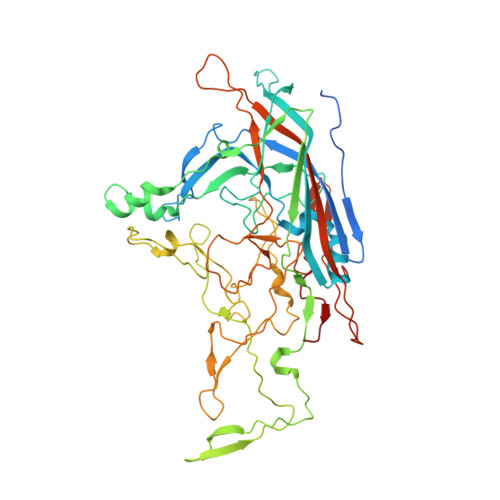
Structure of an enteric pathogen, bovine parvovirus.
Kailasan, S., Halder, S., Gurda, B., Bladek, H., Chipman, P.R., McKenna, R., Brown, K., Agbandje-McKenna, M.(2015) J Virol 89: 2603-2614
- PubMed: 25520501
- DOI: https://doi.org/10.1128/JVI.03157-14
- Primary Citation of Related Structures:
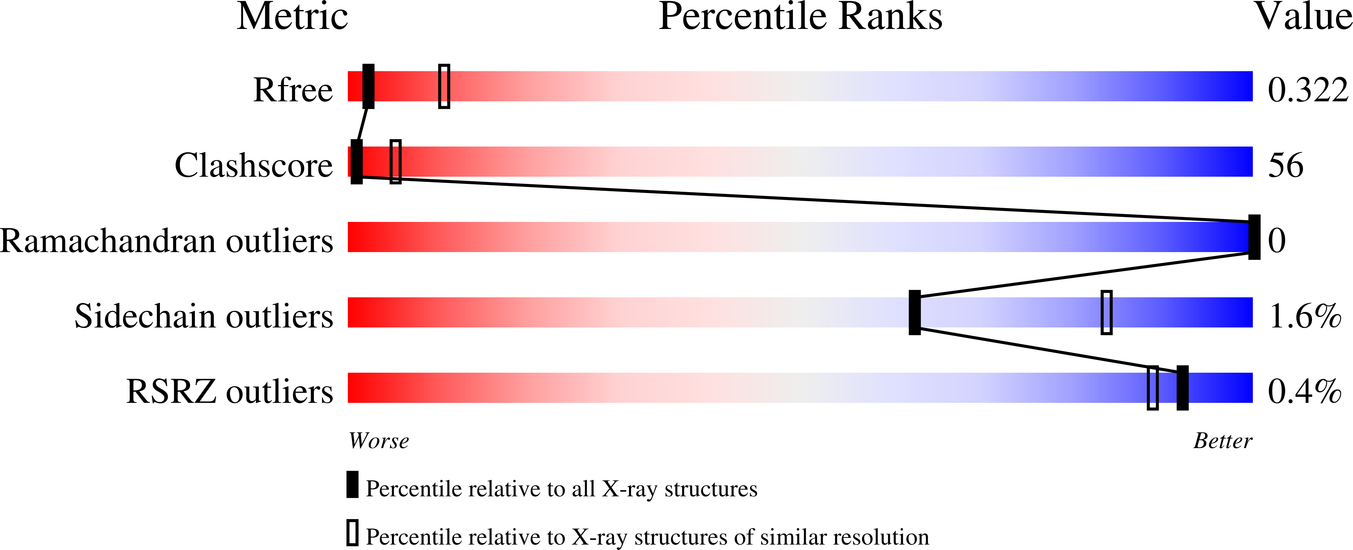
4QC8 - PubMed Abstract:
Bovine parvovirus (BPV), the causative agent of respiratory and gastrointestinal disease in cows, is the type member of the Bocaparvovirus genus of the Parvoviridae family. Toward efforts to obtain a template for the development of vaccines and small-molecule inhibitors for this pathogen, the structure of the BPV capsid, assembled from the major capsid viral protein 2 (VP2), was determined using X-ray crystallography as well as cryo-electron microscopy and three-dimensional image reconstruction (cryo-reconstruction) to 3.2- and 8.8-Å resolutions, respectively. The VP2 region ordered in the crystal structure, from residues 39 to 536, conserves the parvoviral eight-stranded jellyroll motif and an αA helix. The BPV capsid displays common parvovirus features: a channel at and depressions surrounding the 5-fold axes and protrusions surrounding the 3-fold axes. However, rather than a depression centered at the 2-fold axes, a raised surface loop divides this feature in BPV. Additional observed density in the capsid interior in the cryo-reconstructed map, compared to the crystal structure, is interpreted as 10 additional N-terminal residues, residues 29 to 38, that radially extend the channel under the 5-fold axis, as observed for human bocavirus 1 (HBoV1). Surface loops of various lengths and conformations extend from the core jellyroll motif of VP2. These loops confer the unique surface topology of the BPV capsid, making it strikingly different from HBoV1 as well as the type members of other Parvovirinae genera for which structures have been determined. For the type members, regions structurally analogous to those decorating the BPV capsid surface serve as determinants of receptor recognition, tissue and host tropism, pathogenicity, and antigenicity.
Organizational Affiliation:
Department of Biochemistry and Molecular Biology and the McKnight Brain Institute, University of Florida, Gainesville, Florida, USA.