4Q56

Structure of Helix aspersa agglutinin with natural glycosylation and N-acetyl-alpha-D-galactosamine (GalNAc)


Experimental Data Snapshot

  • Method: X-RAY DIFFRACTION
  • Resolution: 1.38 Å
  • R-Value Free: 0.168 
  • R-Value Work: 0.137 
  • R-Value Observed: 0.138 

wwPDB Validation   3D Report Full Report


Ligand Structure Quality Assessment 


This is version 3.1 of the entry. See complete history


Literature

Structural studies of Helix aspersa agglutinin complexed with GalNAc: A lectin that serves as a diagnostic tool.

Pietrzyk, A.J.Bujacz, A.Mak, P.Potempa, B.Niedziela, T.

(2015) Int J Biol Macromol 81: 1059-1068

  • DOI: https://doi.org/10.1016/j.ijbiomac.2015.09.044
  • Primary Citation of Related Structures:  
    4Q56

  • PubMed Abstract: 

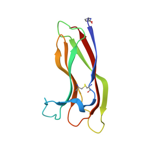
    Lectins belong to a differentiated group of proteins known to possess sugar-binding properties. Due to this fact, they are interesting research targets in medical diagnostics. Helix aspersa agglutinin (HAA) is a lectin that recognizes the epitopes containing α-d-N-acetylgalactosamine (GalNAc), which is present at the surface of metastatic cancer cells. Although several reports have already described the use of HAA as a diagnostic tool, this protein was not characterized on the molecular level. Here, we present for the first time the structural information about lectin isolated from mucus of Helix aspersa (garden snail). The amino acid sequence of this agglutinin was determined by Edman degradation and tertiary as well as quaternary structure by X-ray crystallography. The high resolution crystal structure (1.38Å) and MALDI-TOF mass spectrometry analysis provide the detailed information about a large part of the HAA natural glycan chain. The topology of the GalNAc binding cleft and interaction with lectin are very well defined in the structure and fully confirmed by STD HSQC NMR spectroscopy. Together, this provides structural clues regarding HAA specificity and opens possibilities to rational modifications of this important diagnostic tool.


  • Organizational Affiliation

    Institute of Technical Biochemistry, Faculty of Biotechnology and Food Sciences, Lodz University of Technology, Stefanowskiego 4/10, Lodz 90-924, Poland.


Macromolecules
Find similar proteins by:  (by identity cutoff)  |  3D Structure
Entity ID: 1
MoleculeChains Sequence LengthOrganismDetailsImage
Helix aspersa agglutinin (HAA)101Cornu aspersumMutation(s): 0 
UniProt
Find proteins for A0A0R4I963 (Cornu aspersum)
Explore A0A0R4I963 
Go to UniProtKB:  A0A0R4I963
Entity Groups  
Sequence Clusters30% Identity50% Identity70% Identity90% Identity95% Identity100% Identity
UniProt GroupA0A0R4I963
Sequence Annotations
Expand
  • Reference Sequence
Oligosaccharides

Help

Entity ID: 2
MoleculeChains Length2D Diagram Glycosylation3D Interactions
alpha-L-fucopyranose-(1-2)-beta-D-galactopyranose-(1-4)-alpha-L-fucopyranose-(1-3)-[beta-D-mannopyranose-(1-4)-2-acetamido-2-deoxy-beta-D-glucopyranose-(1-4)]2-acetamido-2-deoxy-beta-D-glucopyranose
B
6N-Glycosylation
Glycosylation Resources
GlyTouCan:  G52988LW
GlyCosmos:  G52988LW
Small Molecules
Modified Residues  1 Unique
IDChains TypeFormula2D DiagramParent
PCA
Query on PCA
A
L-PEPTIDE LINKINGC5 H7 N O3GLN
Experimental Data & Validation

Experimental Data

  • Method: X-RAY DIFFRACTION
  • Resolution: 1.38 Å
  • R-Value Free: 0.168 
  • R-Value Work: 0.137 
  • R-Value Observed: 0.138 
  • Space Group: H 3 2
Unit Cell:
Length ( Å )Angle ( ˚ )
a = 47.9α = 90
b = 47.9β = 90
c = 281.53γ = 120
Software Package:
Software NamePurpose
XDSdata reduction
XDSdata scaling
PHASERphasing
REFMACrefinement

Structure Validation

View Full Validation Report


Ligand Structure Quality Assessment 


Entry History 

Deposition Data

Revision History  (Full details and data files)

  • Version 1.0: 2015-10-14
    Type: Initial release
  • Version 1.1: 2015-10-28
    Changes: Database references
  • Version 1.2: 2018-03-07
    Changes: Data collection
  • Version 2.0: 2019-12-25
    Changes: Data collection, Derived calculations, Polymer sequence
  • Version 3.0: 2020-07-29
    Type: Remediation
    Reason: Carbohydrate remediation
    Changes: Atomic model, Data collection, Derived calculations, Structure summary
  • Version 3.1: 2023-09-20
    Changes: Data collection, Database references, Derived calculations, Refinement description, Structure summary