4PI9

Crystal structure of S. Aureus Autolysin E in complex with muropeptide NAM-L-ALA-D-iGLU


Experimental Data Snapshot

  • Method: X-RAY DIFFRACTION
  • Resolution: 1.48 Å
  • R-Value Free: 0.209 
  • R-Value Work: 0.177 
  • R-Value Observed: 0.179 

wwPDB Validation   3D Report Full Report


Ligand Structure Quality Assessment 


This is version 1.3 of the entry. See complete history


Literature

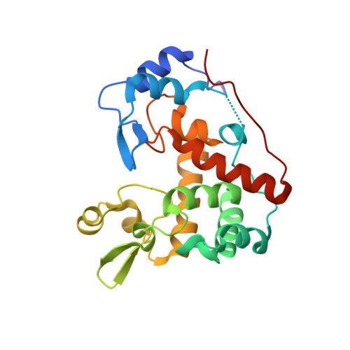
The mechanism behind the selection of two different cleavage sites in NAG-NAM polymers.

Mihelic, M.Vlahovicek-Kahlina, K.Renko, M.Mesnage, S.Dobersek, A.Taler-Vercic, A.Jakas, A.Turk, D.

(2017) IUCrJ 4: 185-198

  • DOI: https://doi.org/10.1107/S2052252517000367
  • Primary Citation of Related Structures:  
    4PI9

  • PubMed Abstract: 

    Peptidoglycan is a giant molecule that forms the cell wall that surrounds bacterial cells. It is composed of alternating N -acetylglucosamine (NAG) and N -acetylmuramic acid (NAM) residues connected by β-(1,4)-glycosidic bonds and cross-linked with short polypeptide chains. Owing to the increasing antibiotic resistance against drugs targeting peptidoglycan synthesis, studies of enzymes involved in the degradation of peptidoglycan, such as N -acetylglucos-aminidases, may expose new, valuable drug targets. The scientific challenge addressed here is how lysozymes, muramidases which are likely to be the most studied enzymes ever, and bacterial N -acetylglucosaminidases discriminate between two glycosidic bonds that are different in sequence yet chemically equivalent in the same NAG-NAM polymers. In spite of more than fifty years of structural studies of lysozyme, it is still not known how the enzyme selects the bond to be cleaved. Using macromolecular crystallography, chemical synthesis and molecular modelling, this study explains how these two groups of enzymes based on an equivalent structural core exhibit a difference in selectivity. The crystal structures of Staphylococcus aureus N -acetylglucosaminidase autolysin E (AtlE) alone and in complex with fragments of peptidoglycan revealed that N -acetylglucosaminidases and muramidases approach the substrate at alternate glycosidic bond positions from opposite sides. The recognition pocket for NAM residues in the active site of N -acetylglucosaminidases may make them a suitable drug target.


  • Organizational Affiliation

    Department of Biochemistry and Molecular and Structural Biology, Jozef Stefan Institute, Jamova 39, 1000 Ljubljana, Slovenia; Centre of Excellence for Integrated Approaches in Chemistry and Biology of Proteins, Jamova 39, 1000 Ljubljana, Slovenia.


Macromolecules
Find similar proteins by:  (by identity cutoff)  |  3D Structure
Entity ID: 1
MoleculeChains Sequence LengthOrganismDetailsImage
Autolysin E228Staphylococcus aureus subsp. aureus Mu50Mutation(s): 0 
Gene Names: SAV2307
UniProt
Find proteins for A0A0H3JT72 (Staphylococcus aureus (strain Mu50 / ATCC 700699))
Explore A0A0H3JT72 
Go to UniProtKB:  A0A0H3JT72
Entity Groups  
Sequence Clusters30% Identity50% Identity70% Identity90% Identity95% Identity100% Identity
UniProt GroupA0A0H3JT72
Sequence Annotations
Expand
  • Reference Sequence
Small Molecules
Ligands 3 Unique
IDChains Name / Formula / InChI Key2D Diagram3D Interactions
3LT
Query on 3LT

Download Ideal Coordinates CCD File 
L [auth A](4R)-4-[[(2S)-2-[[(2R)-2-[(2R,3S,4R,5R,6R)-5-acetamido-2-(hydroxymethyl)-3,6-bis(oxidanyl)oxan-4-yl]oxypropanoyl]amino]propanoyl]amino]-5-azanyl-5-oxidanylidene-pentanoic acid
C19 H32 N4 O11
BSOQXXWZTUDTEL-ZUYCGGNHSA-N
SO4
Query on SO4

Download Ideal Coordinates CCD File 
B [auth A],
C [auth A],
D [auth A]
SULFATE ION
O4 S
QAOWNCQODCNURD-UHFFFAOYSA-L
CL
Query on CL

Download Ideal Coordinates CCD File 
E [auth A]
F [auth A]
G [auth A]
H [auth A]
I [auth A]
E [auth A],
F [auth A],
G [auth A],
H [auth A],
I [auth A],
J [auth A],
K [auth A]
CHLORIDE ION
Cl
VEXZGXHMUGYJMC-UHFFFAOYSA-M
Experimental Data & Validation

Experimental Data

  • Method: X-RAY DIFFRACTION
  • Resolution: 1.48 Å
  • R-Value Free: 0.209 
  • R-Value Work: 0.177 
  • R-Value Observed: 0.179 
  • Space Group: P 21 21 21
Unit Cell:
Length ( Å )Angle ( ˚ )
a = 45.626α = 90
b = 69.312β = 90
c = 73.424γ = 90
Software Package:
Software NamePurpose
REFMACrefinement
PDB_EXTRACTdata extraction
HKL-2000data reduction
HKL-2000data scaling

Structure Validation

View Full Validation Report


Ligand Structure Quality Assessment 


Entry History 

Deposition Data

Revision History  (Full details and data files)

  • Version 1.0: 2015-10-14
    Type: Initial release
  • Version 1.1: 2017-03-08
    Changes: Database references
  • Version 1.2: 2018-04-18
    Changes: Data collection, Database references
  • Version 1.3: 2023-12-27
    Changes: Advisory, Data collection, Database references