4P9O

Complex of Voltage-gated ion channel in a the presence of channel blocking compound


Experimental Data Snapshot

  • Method: X-RAY DIFFRACTION
  • Resolution: 2.89 Å
  • R-Value Free: 0.251 
  • R-Value Work: 0.214 
  • R-Value Observed: 0.216 

wwPDB Validation   3D Report Full Report


Ligand Structure Quality Assessment 


This is version 2.2 of the entry. See complete history


Literature

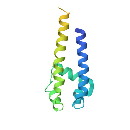
Prokaryotic NavMs channel as a structural and functional model for eukaryotic sodium channel antagonism.

Bagneris, C.DeCaen, P.G.Naylor, C.E.Pryde, D.C.Nobeli, I.Clapham, D.E.Wallace, B.A.

(2014) Proc Natl Acad Sci U S A 111: 8428-8433

  • DOI: https://doi.org/10.1073/pnas.1406855111
  • Primary Citation of Related Structures:  
    4CBC, 4OXS, 4P2Z, 4P30, 4P9O, 4P9P, 4PA3, 4PA4, 4PA6, 4PA7, 4PA9

  • PubMed Abstract: 

    Voltage-gated sodium channels are important targets for the development of pharmaceutical drugs, because mutations in different human sodium channel isoforms have causal relationships with a range of neurological and cardiovascular diseases. In this study, functional electrophysiological studies show that the prokaryotic sodium channel from Magnetococcus marinus (NavMs) binds and is inhibited by eukaryotic sodium channel blockers in a manner similar to the human Nav1.1 channel, despite millions of years of divergent evolution between the two types of channels. Crystal complexes of the NavMs pore with several brominated blocker compounds depict a common antagonist binding site in the cavity, adjacent to lipid-facing fenestrations proposed to be the portals for drug entry. In silico docking studies indicate the full extent of the blocker binding site, and electrophysiology studies of NavMs channels with mutations at adjacent residues validate the location. These results suggest that the NavMs channel can be a valuable tool for screening and rational design of human drugs.


  • Organizational Affiliation

    Institute of Structural and Molecular Biology, School of Biological Sciences, Birkbeck College, University of London, London WC1E 7HX, United Kingdom;


Macromolecules
Find similar proteins by:  (by identity cutoff)  |  3D Structure
Entity ID: 1
MoleculeChains Sequence LengthOrganismDetailsImage
Ion transport protein
A, B, C, D
149Magnetococcus marinus MC-1Mutation(s): 0 
Gene Names: Mmc1_0798
Membrane Entity: Yes 
UniProt
Find proteins for A0L5S6 (Magnetococcus marinus (strain ATCC BAA-1437 / JCM 17883 / MC-1))
Explore A0L5S6 
Go to UniProtKB:  A0L5S6
Entity Groups  
Sequence Clusters30% Identity50% Identity70% Identity90% Identity95% Identity100% Identity
UniProt GroupA0L5S6
Sequence Annotations
Expand
  • Reference Sequence
Small Molecules
Ligands 2 Unique
IDChains Name / Formula / InChI Key2D Diagram3D Interactions
2CV
Query on 2CV

Download Ideal Coordinates CCD File 
E [auth A]
F [auth A]
H [auth B]
I [auth B]
K [auth C]
E [auth A],
F [auth A],
H [auth B],
I [auth B],
K [auth C],
M [auth D]
HEGA-10
C18 H37 N O7
ITEIKACYSCODFV-ATLSCFEFSA-N
BR
Query on BR

Download Ideal Coordinates CCD File 
G [auth B],
J [auth B],
L [auth D],
N [auth D]
BROMIDE ION
Br
CPELXLSAUQHCOX-UHFFFAOYSA-M
Experimental Data & Validation

Experimental Data

  • Method: X-RAY DIFFRACTION
  • Resolution: 2.89 Å
  • R-Value Free: 0.251 
  • R-Value Work: 0.214 
  • R-Value Observed: 0.216 
  • Space Group: C 2 2 21
Unit Cell:
Length ( Å )Angle ( ˚ )
a = 80.42α = 90
b = 328.37β = 90
c = 80.39γ = 90
Software Package:
Software NamePurpose
BUSTERrefinement

Structure Validation

View Full Validation Report


Ligand Structure Quality Assessment 


Entry History & Funding Information

Deposition Data


Funding OrganizationLocationGrant Number
Biotechnology and Biological Sciences Research Council (BBSRC)United KingdomBB/H02070X
Biotechnology and Biological Sciences Research Council (BBSRC)United KingdomBB/J020702

Revision History  (Full details and data files)

  • Version 1.0: 2014-06-04
    Type: Initial release
  • Version 1.1: 2014-08-06
    Changes: Database references
  • Version 2.0: 2017-09-20
    Changes: Atomic model, Author supporting evidence, Data collection, Database references, Derived calculations, Other, Source and taxonomy, Structure summary
  • Version 2.1: 2020-01-01
    Changes: Author supporting evidence
  • Version 2.2: 2023-12-27
    Changes: Data collection, Database references, Refinement description