4P5H

Structure of Clostridium perfringens Enterotoxin with a peptide derived from a modified version of ECL-2 of Claudin 2


Experimental Data Snapshot

  • Method: X-RAY DIFFRACTION
  • Resolution: 3.38 Å
  • R-Value Free: 0.240 
  • R-Value Work: 0.204 
  • R-Value Observed: 0.206 

wwPDB Validation   3D Report Full Report


This is version 1.6 of the entry. See complete history


Literature

Structure of a C. perfringens Enterotoxin Mutant in Complex with a Modified Claudin-2 Extracellular Loop 2.

Yelland, T.S.Naylor, C.E.Bagoban, T.Savva, C.G.Moss, D.S.McClane, B.A.Blasig, I.E.Popoff, M.Basak, A.K.

(2014) J Mol Biol 426: 3134-3147

  • DOI: https://doi.org/10.1016/j.jmb.2014.07.001
  • Primary Citation of Related Structures:  
    3ZIW, 3ZIX, 4P5H

  • PubMed Abstract: 

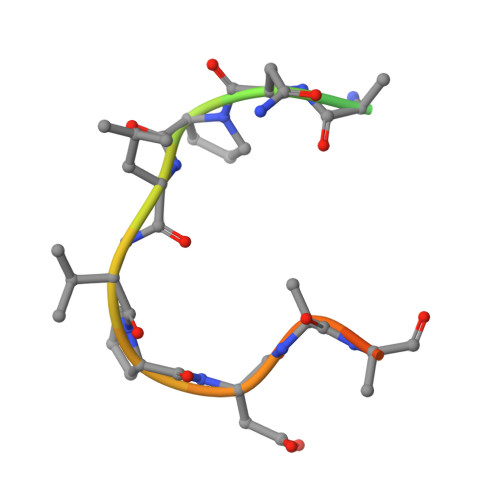
    CPE (Clostridium perfringens enterotoxin) is the major virulence determinant for C. perfringens type-A food poisoning, the second most common bacterial food-borne illness in the UK and USA. After binding to its receptors, which include particular human claudins, the toxin forms pores in the cell membrane. The mature pore apparently contains a hexamer of CPE, claudin and, possibly, occludin. The combination of high binding specificity with cytotoxicity has resulted in CPE being investigated, with some success, as a targeted cytotoxic agent for oncotherapy. In this paper, we present the X-ray crystallographic structure of CPE in complex with a peptide derived from extracellular loop 2 of a modified, CPE-binding Claudin-2, together with high-resolution native and pore-formation mutant structures. Our structure provides the first atomic-resolution data on any part of a claudin molecule and reveals that claudin's CPE-binding fingerprint (NPLVP) is in a tight turn conformation and binds, as expected, in CPE's C-terminal claudin-binding groove. The leucine and valine residues insert into the binding groove while the first residue, asparagine, tethers the peptide via an interaction with CPE's aspartate 225 and the two prolines are required to maintain the tight turn conformation. Understanding the structural basis of the contribution these residues make to binding will aid in engineering CPE to target tumor cells.


  • Organizational Affiliation

    Department of Biological Sciences, Birkbeck College, London WC1E 7HX, UK.


Macromolecules

Find similar proteins by:  Sequence   |   3D Structure  

Entity ID: 1
MoleculeChains Sequence LengthOrganismDetailsImage
Claudin-220Mus musculusMutation(s): 2 
UniProt
Find proteins for O88552 (Mus musculus)
Explore O88552 
Go to UniProtKB:  O88552
Entity Groups  
Sequence Clusters30% Identity50% Identity70% Identity90% Identity95% Identity100% Identity
UniProt GroupO88552
Sequence Annotations
Expand
  • Reference Sequence
Find similar proteins by:  (by identity cutoff)  |  3D Structure
Entity ID: 2
MoleculeChains Sequence LengthOrganismDetailsImage
Heat-labile enterotoxin B chain286Clostridium perfringensMutation(s): 0 
Gene Names: cpe
UniProt
Find proteins for P01558 (Clostridium perfringens)
Explore P01558 
Go to UniProtKB:  P01558
Entity Groups  
Sequence Clusters30% Identity50% Identity70% Identity90% Identity95% Identity100% Identity
UniProt GroupP01558
Sequence Annotations
Expand
  • Reference Sequence
Experimental Data & Validation

Experimental Data

  • Method: X-RAY DIFFRACTION
  • Resolution: 3.38 Å
  • R-Value Free: 0.240 
  • R-Value Work: 0.204 
  • R-Value Observed: 0.206 
  • Space Group: C 1 2 1
Unit Cell:
Length ( Å )Angle ( ˚ )
a = 369.6α = 90
b = 100.26β = 119.74
c = 265.36γ = 90
Software Package:
Software NamePurpose
BUSTERrefinement

Structure Validation

View Full Validation Report


Entry History & Funding Information

Deposition Data


Funding OrganizationLocationGrant Number
Medical Research Council (MRC, United Kingdom)United KingdomG0700051
Wellcome TrustUnited KingdomWT089618MA
National Institutes of Health/National Institute Of Allergy and Infectious Diseases (NIH/NIAID)United StatesR37 AI19844-30

Revision History  (Full details and data files)

  • Version 1.0: 2014-07-16
    Type: Initial release
  • Version 1.1: 2014-07-23
    Changes: Database references
  • Version 1.2: 2014-08-06
    Changes: Other
  • Version 1.3: 2014-10-01
    Changes: Database references
  • Version 1.4: 2017-09-27
    Changes: Author supporting evidence, Database references, Derived calculations, Other, Source and taxonomy
  • Version 1.5: 2019-12-11
    Changes: Author supporting evidence
  • Version 1.6: 2023-09-27
    Changes: Data collection, Database references, Refinement description