4O65

Crystal structure of the cupredoxin domain of amoB from Nitrosocaldus yellowstonii


Experimental Data Snapshot

  • Method: X-RAY DIFFRACTION
  • Resolution: 1.80 Å
  • R-Value Free: 0.229 
  • R-Value Work: 0.181 
  • R-Value Observed: 0.183 

wwPDB Validation   3D Report Full Report


This is version 1.2 of the entry. See complete history


Literature

Structural conservation of the B subunit in the ammonia monooxygenase/particulate methane monooxygenase superfamily.

Lawton, T.J.Ham, J.Sun, T.Rosenzweig, A.C.

(2014) Proteins 82: 2263-2267

  • DOI: https://doi.org/10.1002/prot.24535
  • Primary Citation of Related Structures:  
    4O65

  • PubMed Abstract: 

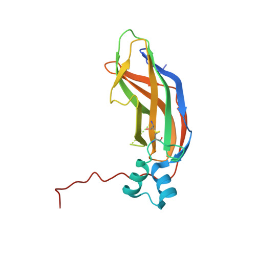
    The ammonia monooxygenase (AMO)/particulate methane monooxygenase (pMMO) superfamily is a diverse group of membrane-bound enzymes of which only pMMO has been characterized on the molecular level. The pMMO active site is believed to reside in the soluble N-terminal region of the pmoB subunit. To understand the degree of structural conservation within this superfamily, the crystal structure of the corresponding domain of an archaeal amoB subunit from Nitrosocaldus yellowstonii has been determined to 1.8 Å resolution. The structure reveals a remarkable conservation of overall fold and copper binding site location as well as several notable differences that may have implications for function and stability.


  • Organizational Affiliation

    Departments of Molecular Biosciences and of Chemistry, Northwestern University, Evanston, Illinois, 60208.


Macromolecules
Find similar proteins by:  (by identity cutoff)  |  3D Structure
Entity ID: 1
MoleculeChains Sequence LengthOrganismDetailsImage
Putative archaeal ammonia monooxygenase subunit B166Candidatus Nitrosocaldus yellowstonensisMutation(s): 0 
Gene Names: amoB
UniProt
Find proteins for B0LKZ4 (Candidatus Nitrosocaldus yellowstonensis)
Explore B0LKZ4 
Go to UniProtKB:  B0LKZ4
Entity Groups  
Sequence Clusters30% Identity50% Identity70% Identity90% Identity95% Identity100% Identity
UniProt GroupB0LKZ4
Sequence Annotations
Expand
  • Reference Sequence
Experimental Data & Validation

Experimental Data

  • Method: X-RAY DIFFRACTION
  • Resolution: 1.80 Å
  • R-Value Free: 0.229 
  • R-Value Work: 0.181 
  • R-Value Observed: 0.183 
  • Space Group: P 43 21 2
Unit Cell:
Length ( Å )Angle ( ˚ )
a = 47.886α = 90
b = 47.886β = 90
c = 192.614γ = 90
Software Package:
Software NamePurpose
HKL-2000data collection
SHARPphasing
REFMACrefinement
HKL-2000data reduction
HKL-2000data scaling

Structure Validation

View Full Validation Report


Entry History 

Deposition Data

Revision History  (Full details and data files)

  • Version 1.0: 2014-04-02
    Type: Initial release
  • Version 1.1: 2014-09-03
    Changes: Database references
  • Version 1.2: 2014-11-12
    Changes: Structure summary