4NHJ

Crystal structure of Klebsiella pneumoniae RstA DNA-binding domain in complex with RstA box


Experimental Data Snapshot

  • Method: X-RAY DIFFRACTION
  • Resolution: 2.70 Å
  • R-Value Free: 0.271 
  • R-Value Work: 0.216 
  • R-Value Observed: 0.222 

wwPDB Validation   3D Report Full Report


This is version 1.2 of the entry. See complete history


Literature

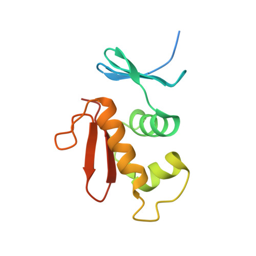
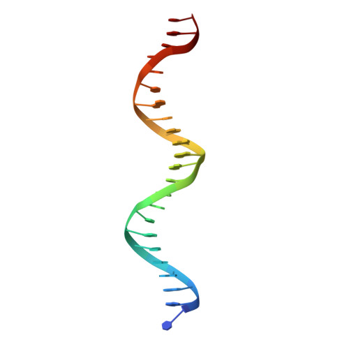
Structural dynamics of the two-component response regulator RstA in recognition of promoter DNA element.

Li, Y.C.Chang, C.K.Chang, C.F.Cheng, Y.H.Fang, P.J.Yu, T.Chen, S.C.Li, Y.C.Hsiao, C.D.Huang, T.H.

(2014) Nucleic Acids Res 42: 8777-8788

  • DOI: https://doi.org/10.1093/nar/gku572
  • Primary Citation of Related Structures:  
    2MLK, 4NHJ, 4NIC

  • PubMed Abstract: 

    The RstA/RstB system is a bacterial two-component regulatory system consisting of the membrane sensor, RstB and its cognate response regulator (RR) RstA. The RstA of Klebsiella pneumoniae (kpRstA) consists of an N-terminal receiver domain (RD, residues 1-119) and a C-terminal DNA-binding domain (DBD, residues 130-236). Phosphorylation of kpRstA induces dimerization, which allows two kpRstA DBDs to bind to a tandem repeat, called the RstA box, and regulate the expression of downstream genes. Here we report the solution and crystal structures of the free kpRstA RD, DBD and DBD/RstA box DNA complex. The structure of the kpRstA DBD/RstA box complex suggests that the two protomers interact with the RstA box in an asymmetric fashion. Equilibrium binding studies further reveal that the two protomers within the kpRstA dimer bind to the RstA box in a sequential manner. Taken together, our results suggest a binding model where dimerization of the kpRstA RDs provides the platform to allow the first kpRstA DBD protomer to anchor protein-DNA interaction, whereas the second protomer plays a key role in ensuring correct recognition of the RstA box.


  • Organizational Affiliation

    Institute of Molecular Biology, Academia Sinica, Taipei 115, Taiwan, ROC Institute of Bioinformatics and Structural Biology, National Tsing Hua University, Hsinchu 300, Taiwan.


Macromolecules

Find similar proteins by:  (by identity cutoff)  |  3D Structure
Entity ID: 1
MoleculeChains Sequence LengthOrganismDetailsImage
DNA-binding transcriptional regulator RstA
A, B
119Klebsiella pneumoniaeMutation(s): 2 
Gene Names: rstAKPN2242_10390KPR_2822
UniProt
Find proteins for A6T8N1 (Klebsiella pneumoniae subsp. pneumoniae (strain ATCC 700721 / MGH 78578))
Explore A6T8N1 
Go to UniProtKB:  A6T8N1
Entity Groups  
Sequence Clusters30% Identity50% Identity70% Identity90% Identity95% Identity100% Identity
UniProt GroupA6T8N1
Sequence Annotations
Expand
  • Reference Sequence

Find similar nucleic acids by:  Sequence   |   3D Structure  

Entity ID: 2
MoleculeChains LengthOrganismImage
5'-D(*GP*GP*TP*TP*GP*TP*AP*CP*AP*TP*TP*CP*CP*GP*TP*TP*AP*CP*TP*CP*CP*CP*T)-3'23N/A
Sequence Annotations
Expand
  • Reference Sequence

Find similar nucleic acids by:  Sequence   |   3D Structure  

Entity ID: 3
MoleculeChains LengthOrganismImage
5'-D(*CP*AP*GP*GP*GP*AP*GP*TP*AP*AP*CP*GP*GP*AP*AP*TP*GP*TP*AP*CP*AP*AP*C)-3'23N/A
Sequence Annotations
Expand
  • Reference Sequence
Experimental Data & Validation

Experimental Data

  • Method: X-RAY DIFFRACTION
  • Resolution: 2.70 Å
  • R-Value Free: 0.271 
  • R-Value Work: 0.216 
  • R-Value Observed: 0.222 
  • Space Group: P 21 21 2
Unit Cell:
Length ( Å )Angle ( ˚ )
a = 159.935α = 90
b = 33.313β = 90
c = 72.779γ = 90
Software Package:
Software NamePurpose
HKL-2000data collection
SOLVEphasing
PHENIXrefinement
HKL-2000data reduction
HKL-2000data scaling

Structure Validation

View Full Validation Report


Entry History 

Deposition Data

Revision History  (Full details and data files)

  • Version 1.0: 2014-07-16
    Type: Initial release
  • Version 1.1: 2014-08-13
    Changes: Database references
  • Version 1.2: 2024-02-28
    Changes: Data collection, Database references