


Crystal structure of (S)-3-hydroxybutylryl-CoA dehydrogenase form the n-butanol sysnthesizing bacterium, Clostridium butyricum
Kim, E.J., Kim, S., Kim, K.J.To be published.
Experimental Data Snapshot
Entity ID: 1 | |||||
|---|---|---|---|---|---|
| Molecule | Chains | Sequence Length | Organism | Details | Image |
| 3-hydroxybutyryl-CoA dehydrogenase | 290 | Clostridium butyricum E4 str. BoNT E BL5262 | Mutation(s): 0 Gene Names: hbd, CLP_3850 EC: 1.1.1.157 | 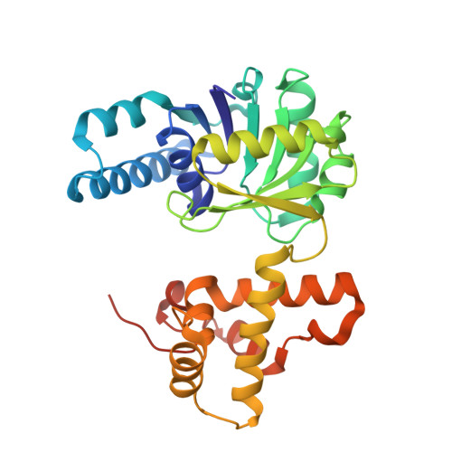 | |
UniProt | |||||
Find proteins for C4IEM5 (Clostridium butyricum E4 str. BoNT E BL5262) Explore C4IEM5 Go to UniProtKB: C4IEM5 | |||||
Entity Groups | |||||
| Sequence Clusters | 30% Identity50% Identity70% Identity90% Identity95% Identity100% Identity | ||||
| UniProt Group | C4IEM5 | ||||
Sequence AnnotationsExpand | |||||
| |||||
| Ligands 1 Unique | |||||
|---|---|---|---|---|---|
| ID | Chains | Name / Formula / InChI Key | 2D Diagram | 3D Interactions | |
| NAD Query on NAD | E [auth A], F [auth B], G [auth C], H [auth D] | NICOTINAMIDE-ADENINE-DINUCLEOTIDE C21 H27 N7 O14 P2 BAWFJGJZGIEFAR-NNYOXOHSSA-N |  | ||
| Length ( Å ) | Angle ( ˚ ) |
|---|---|
| a = 148.45 | α = 90 |
| b = 148.45 | β = 90 |
| c = 201.63 | γ = 120 |
| Software Name | Purpose |
|---|---|
| DENZO | data reduction |
| SCALEPACK | data scaling |
| MOLREP | phasing |
| REFMAC | refinement |
| PDB_EXTRACT | data extraction |
RCSB PDB (citation) is hosted by
RCSB PDB is a member of the

