4K30

Structure of the N-acetyltransferase domain of human N-acetylglutamate synthase


Experimental Data Snapshot

  • Method: X-RAY DIFFRACTION
  • Resolution: 2.10 Å
  • R-Value Free: 0.244 
  • R-Value Work: 0.185 
  • R-Value Observed: 0.187 

wwPDB Validation   3D Report Full Report


Ligand Structure Quality Assessment 


This is version 1.2 of the entry. See complete history


Literature

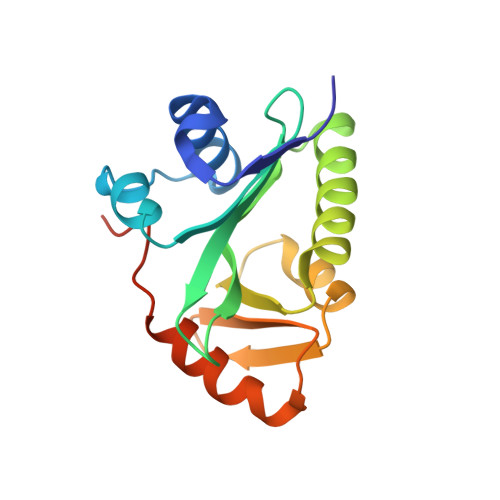
Crystal structure of the N-acetyltransferase domain of human N-acetyl-L-glutamate synthase in complex with N-acetyl-L-glutamate provides insights into its catalytic and regulatory mechanisms.

Zhao, G.Jin, Z.Allewell, N.M.Tuchman, M.Shi, D.

(2013) PLoS One 8: e70369-e70369

  • DOI: https://doi.org/10.1371/journal.pone.0070369
  • Primary Citation of Related Structures:  
    4K30

  • PubMed Abstract: 

    N-acetylglutamate synthase (NAGS) catalyzes the conversion of AcCoA and L-glutamate to CoA and N-acetyl-L-glutamate (NAG), an obligate cofactor for carbamyl phosphate synthetase I (CPSI) in the urea cycle. NAGS deficiency results in elevated levels of plasma ammonia which is neurotoxic. We report herein the first crystal structure of human NAGS, that of the catalytic N-acetyltransferase (hNAT) domain with N-acetyl-L-glutamate bound at 2.1 Å resolution. Functional studies indicate that the hNAT domain retains catalytic activity in the absence of the amino acid kinase (AAK) domain. Instead, the major functions of the AAK domain appear to be providing a binding site for the allosteric activator, L-arginine, and an N-terminal proline-rich motif that is likely to function in signal transduction to CPS1. Crystalline hNAT forms a dimer similar to the NAT-NAT dimers that form in crystals of bifunctional N-acetylglutamate synthase/kinase (NAGS/K) from Maricaulis maris and also exists as a dimer in solution. The structure of the NAG binding site, in combination with mutagenesis studies, provide insights into the catalytic mechanism. We also show that native NAGS from human and mouse exists in tetrameric form, similar to those of bifunctional NAGS/K.


  • Organizational Affiliation

    Center for Genetic Medicine Research and Department of Integrative Systems Biology, Children's National Medical Center, The George Washington University, Washington, DC, United States of America.


Macromolecules
Find similar proteins by:  (by identity cutoff)  |  3D Structure
Entity ID: 1
MoleculeChains Sequence LengthOrganismDetailsImage
N-acetylglutamate synthase, mitochondrialA,
B,
C [auth X],
D [auth Y]
160Homo sapiensMutation(s): 0 
Gene Names: NAGS
EC: 2.3.1.1
UniProt & NIH Common Fund Data Resources
Find proteins for Q8N159 (Homo sapiens)
Explore Q8N159 
Go to UniProtKB:  Q8N159
PHAROS:  Q8N159
GTEx:  ENSG00000161653 
Entity Groups  
Sequence Clusters30% Identity50% Identity70% Identity90% Identity95% Identity100% Identity
UniProt GroupQ8N159
Sequence Annotations
Expand
  • Reference Sequence
Experimental Data & Validation

Experimental Data

  • Method: X-RAY DIFFRACTION
  • Resolution: 2.10 Å
  • R-Value Free: 0.244 
  • R-Value Work: 0.185 
  • R-Value Observed: 0.187 
  • Space Group: P 43 21 2
Unit Cell:
Length ( Å )Angle ( ˚ )
a = 116.141α = 90
b = 116.141β = 90
c = 109.742γ = 90
Software Package:
Software NamePurpose
SCALEPACKdata scaling
PHASERphasing
PHENIXrefinement
PDB_EXTRACTdata extraction
CBASSdata collection
DENZOdata reduction
HKL-2000data scaling

Structure Validation

View Full Validation Report


Ligand Structure Quality Assessment 


Entry History 

Deposition Data

Revision History  (Full details and data files)

  • Version 1.0: 2013-08-14
    Type: Initial release
  • Version 1.1: 2017-11-15
    Changes: Refinement description, Structure summary
  • Version 1.2: 2023-09-20
    Changes: Data collection, Database references, Derived calculations, Refinement description