4IOU

Crystal structure of the HIV-1 Vif binding, catalytically active domain of APOBEC3F


Experimental Data Snapshot

  • Method: X-RAY DIFFRACTION
  • Resolution: 2.75 Å
  • R-Value Free: 0.233 
  • R-Value Work: 0.193 
  • R-Value Observed: 0.195 

wwPDB Validation   3D Report Full Report


This is version 1.4 of the entry. See complete history


Literature

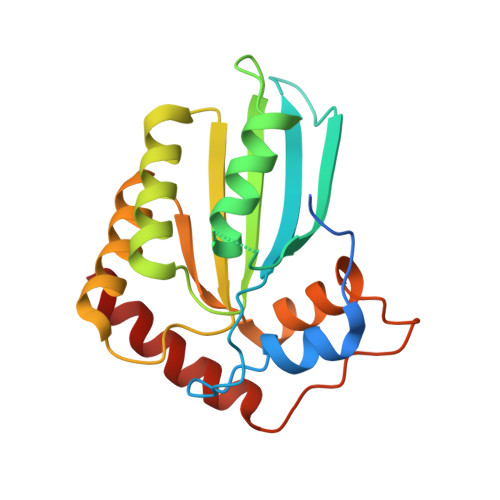
Crystal Structure of the DNA Cytosine Deaminase APOBEC3F: The Catalytically Active and HIV-1 Vif-Binding Domain.

Bohn, M.F.Shandilya, S.M.Albin, J.S.Kouno, T.Anderson, B.D.McDougle, R.M.Carpenter, M.A.Rathore, A.Evans, L.Davis, A.N.Zhang, J.Lu, Y.Somasundaran, M.Matsuo, H.Harris, R.S.Schiffer, C.A.

(2013) Structure 21: 1042-1050

  • DOI: https://doi.org/10.1016/j.str.2013.04.010
  • Primary Citation of Related Structures:  
    4IOU

  • PubMed Abstract: 

    Human APOBEC3F is an antiretroviral single-strand DNA cytosine deaminase, susceptible to degradation by the HIV-1 protein Vif. In this study the crystal structure of the HIV Vif binding, catalytically active, C-terminal domain of APOBEC3F (A3F-CTD) was determined. The A3F-CTD shares structural motifs with portions of APOBEC3G-CTD, APOBEC3C, and APOBEC2. Residues identified to be critical for Vif-dependent degradation of APOBEC3F all fit within a predominantly negatively charged contiguous region on the surface of A3F-CTD. Specific sequence motifs, previously shown to play a role in Vif susceptibility and virion encapsidation, are conserved across APOBEC3s and between APOBEC3s and HIV-1 Vif. In this structure these motifs pack against each other at intermolecular interfaces, providing potential insights both into APOBEC3 oligomerization and Vif interactions.


  • Organizational Affiliation

    Department of Biochemistry and Molecular Pharmacology, University of Massachusetts Medical School, Worcester, MA 01605, USA.


Macromolecules
Find similar proteins by:  (by identity cutoff)  |  3D Structure
Entity ID: 1
MoleculeChains Sequence LengthOrganismDetailsImage
DNA dC->dU-editing enzyme APOBEC-3F
A, B, C, D
199Homo sapiensMutation(s): 11 
Gene Names: APOBEC3F
EC: 3.5.4
UniProt & NIH Common Fund Data Resources
Find proteins for Q8IUX4 (Homo sapiens)
Explore Q8IUX4 
Go to UniProtKB:  Q8IUX4
PHAROS:  Q8IUX4
GTEx:  ENSG00000128394 
Entity Groups  
Sequence Clusters30% Identity50% Identity70% Identity90% Identity95% Identity100% Identity
UniProt GroupQ8IUX4
Sequence Annotations
Expand
  • Reference Sequence
Experimental Data & Validation

Experimental Data

  • Method: X-RAY DIFFRACTION
  • Resolution: 2.75 Å
  • R-Value Free: 0.233 
  • R-Value Work: 0.193 
  • R-Value Observed: 0.195 
  • Space Group: P 1
Unit Cell:
Length ( Å )Angle ( ˚ )
a = 50.79α = 110.75
b = 66.91β = 94.41
c = 75.64γ = 108.83
Software Package:
Software NamePurpose
PHENIXrefinement
PHASERphasing
REFMACrefinement
HKL-2000data reduction
HKL-2000data scaling
Locallydata collection

Structure Validation

View Full Validation Report


Entry History 

Deposition Data

Revision History  (Full details and data files)

  • Version 1.0: 2013-05-29
    Type: Initial release
  • Version 1.1: 2013-06-19
    Changes: Database references, Other, Refinement description
  • Version 1.2: 2013-07-03
    Changes: Database references
  • Version 1.3: 2019-07-17
    Changes: Data collection, Refinement description
  • Version 1.4: 2023-09-20
    Changes: Data collection, Database references, Derived calculations, Refinement description