4IFE

Crystal Structure of the large terminase subunit gp2 of bacterial virus Sf6 complexed with ATP


Experimental Data Snapshot

  • Method: X-RAY DIFFRACTION
  • Resolution: 3.05 Å
  • R-Value Free: 0.279 
  • R-Value Work: 0.208 
  • R-Value Observed: 0.212 

wwPDB Validation   3D Report Full Report


Ligand Structure Quality Assessment 


This is version 1.3 of the entry. See complete history


Literature

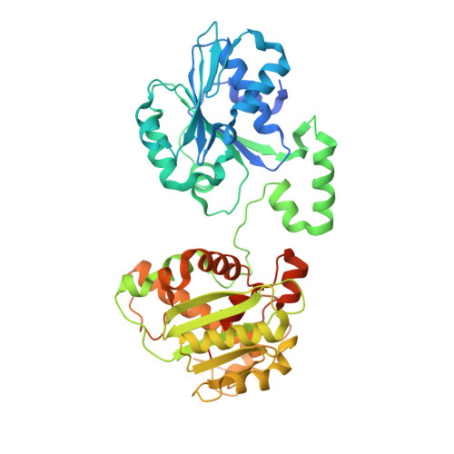
Structures of the phage Sf6 large terminase provide new insights into DNA translocation and cleavage.

Zhao, H.Christensen, T.E.Kamau, Y.N.Tang, L.

(2013) Proc Natl Acad Sci U S A 110: 8075-8080

  • DOI: https://doi.org/10.1073/pnas.1301133110
  • Primary Citation of Related Structures:  
    4IDH, 4IEE, 4IEI, 4IFE

  • PubMed Abstract: 

    Many DNA viruses use powerful molecular motors to cleave concatemeric viral DNA into genome-length units and package them into preformed procapsid powered by ATP hydrolysis. Here we report the structures of the DNA-packaging motor gp2 of bacteriophage Sf6, which reveal a unique clade of RecA-like ATPase domain and an RNase H-like nuclease domain tethered by a regulatory linker domain, exhibiting a strikingly distinct domain arrangement. The gp2 structures complexed with nucleotides reveal, at the atomic detail, the catalytic center embraced by the ATPase domain and the linker domain. The gp2 nuclease activity is modulated by the ATPase domain and is stimulated by ATP. An extended DNA-binding surface is formed by the linker domain and the nuclease domain. These results suggest a unique mechanism for translation of chemical reaction into physical motion of DNA and provide insights into coordination of DNA translocation and cleavage in a viral DNA-packaging motor, which may be achieved via linker-domain-mediated interdomain communication driven by ATP hydrolysis.


  • Organizational Affiliation

    Department of Molecular Biosciences, University of Kansas, Lawrence, KS 66045, USA.


Macromolecules
Find similar proteins by:  (by identity cutoff)  |  3D Structure
Entity ID: 1
MoleculeChains Sequence LengthOrganismDetailsImage
Gene 2 protein490Lederbergvirus Sf6Mutation(s): 0 
UniProt
Find proteins for Q716H3 (Shigella phage Sf6)
Explore Q716H3 
Go to UniProtKB:  Q716H3
Entity Groups  
Sequence Clusters30% Identity50% Identity70% Identity90% Identity95% Identity100% Identity
UniProt GroupQ716H3
Sequence Annotations
Expand
  • Reference Sequence
Small Molecules
Ligands 1 Unique
IDChains Name / Formula / InChI Key2D Diagram3D Interactions
ATP
Query on ATP

Download Ideal Coordinates CCD File 
B [auth A]ADENOSINE-5'-TRIPHOSPHATE
C10 H16 N5 O13 P3
ZKHQWZAMYRWXGA-KQYNXXCUSA-N
Experimental Data & Validation

Experimental Data

  • Method: X-RAY DIFFRACTION
  • Resolution: 3.05 Å
  • R-Value Free: 0.279 
  • R-Value Work: 0.208 
  • R-Value Observed: 0.212 
  • Space Group: P 21 21 21
Unit Cell:
Length ( Å )Angle ( ˚ )
a = 58.176α = 90
b = 63.396β = 90
c = 151.212γ = 90
Software Package:
Software NamePurpose
PHENIXrefinement

Structure Validation

View Full Validation Report


Ligand Structure Quality Assessment 


Entry History 

Deposition Data

Revision History  (Full details and data files)

  • Version 1.0: 2013-05-01
    Type: Initial release
  • Version 1.1: 2013-05-15
    Changes: Database references
  • Version 1.2: 2013-05-29
    Changes: Database references
  • Version 1.3: 2023-09-20
    Changes: Data collection, Database references, Derived calculations, Refinement description