4I6X

Crystal Structure of Non-catalyic Domain of Protein Disulfide Isomerase-related (PDIr) Protein


Experimental Data Snapshot

  • Method: X-RAY DIFFRACTION
  • Resolution: 1.50 Å
  • R-Value Free: 0.237 
  • R-Value Work: 0.191 
  • R-Value Observed: 0.193 

wwPDB Validation   3D Report Full Report


This is version 1.1 of the entry. See complete history


Literature

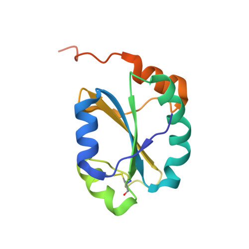
Structure of the Non-Catalytic Domain of the Protein Disulfide Isomerase-Related Protein (PDIR) Reveals Function in Protein Binding.

Vinaik, R.Kozlov, G.Gehring, K.

(2013) PLoS One 8: e62021-e62021

  • DOI: https://doi.org/10.1371/journal.pone.0062021
  • Primary Citation of Related Structures:  
    4I6X

  • PubMed Abstract: 

    Protein disulfide isomerases comprise a large family of enzymes responsible for catalyzing the proper oxidation and folding of newly synthesized proteins in the endoplasmic reticulum (ER). Protein disulfide isomerase-related (PDIR) protein (also known as PDIA5) is a specialized member that participates in the folding of α1-antitrypsin and N-linked glycoproteins. Here, the crystal structure of the non-catalytic domain of PDIR was determined to 1.5 Å resolution. The structure adopts a thioredoxin-like fold stabilized by a structural disulfide bridge with a positively charged binding surface for interactions with the ER chaperones, calreticulin and ERp72. Crystal contacts between molecules potentially mimic the interactions of PDIR with misfolded substrate proteins. The results suggest that the non-catalytic domain of PDIR plays a key role in the recognition of protein partners and substrates.


  • Organizational Affiliation

    Department of Biochemistry, Groupe de Recherche Axé sur la Structure des Protéines, McGill University, Montréal, Québec, Canada.


Macromolecules
Find similar proteins by:  (by identity cutoff)  |  3D Structure
Entity ID: 1
MoleculeChains Sequence LengthOrganismDetailsImage
Protein disulfide-isomerase A5131Homo sapiensMutation(s): 0 
Gene Names: PDIA5PDIR
EC: 5.3.4.1
UniProt & NIH Common Fund Data Resources
Find proteins for Q14554 (Homo sapiens)
Explore Q14554 
Go to UniProtKB:  Q14554
PHAROS:  Q14554
GTEx:  ENSG00000065485 
Entity Groups  
Sequence Clusters30% Identity50% Identity70% Identity90% Identity95% Identity100% Identity
UniProt GroupQ14554
Sequence Annotations
Expand
  • Reference Sequence
Experimental Data & Validation

Experimental Data

  • Method: X-RAY DIFFRACTION
  • Resolution: 1.50 Å
  • R-Value Free: 0.237 
  • R-Value Work: 0.191 
  • R-Value Observed: 0.193 
  • Space Group: P 21 21 21
Unit Cell:
Length ( Å )Angle ( ˚ )
a = 34.686α = 90
b = 41.265β = 90
c = 90.893γ = 90
Software Package:
Software NamePurpose
ADSCdata collection
PHENIXmodel building
REFMACrefinement
HKL-2000data reduction
HKL-2000data scaling
PHENIXphasing

Structure Validation

View Full Validation Report


Entry History 

Deposition Data

Revision History  (Full details and data files)

  • Version 1.0: 2013-04-03
    Type: Initial release
  • Version 1.1: 2013-05-08
    Changes: Database references