


Crystal structure of a hypothetical leucine rich repeat protein (EUBVEN_01088) from Eubacterium ventriosum ATCC 27560 at 2.50 A resolution (CASP Target)
Joint Center for Structural Genomics (JCSG)To be published.
Experimental Data Snapshot
wwPDB Validation 3D Report Full Report

Entity ID: 1 | |||||
|---|---|---|---|---|---|
| Molecule | Chains | Sequence Length | Organism | Details | Image |
| Hypothetical leucine rich repeat protein | 379 | Eubacterium ventriosum ATCC 27560 | Mutation(s): 0 Gene Names: EUBVEN_01088 | 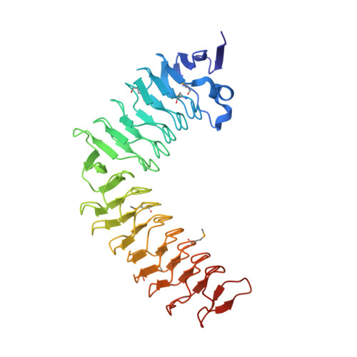 | |
UniProt | |||||
Find proteins for A5Z5V6 (Eubacterium ventriosum ATCC 27560) Explore A5Z5V6 Go to UniProtKB: A5Z5V6 | |||||
Entity Groups | |||||
| Sequence Clusters | 30% Identity50% Identity70% Identity90% Identity95% Identity100% Identity | ||||
| UniProt Group | A5Z5V6 | ||||
Sequence AnnotationsExpand | |||||
| |||||
| Ligands 2 Unique | |||||
|---|---|---|---|---|---|
| ID | Chains | Name / Formula / InChI Key | 2D Diagram | 3D Interactions | |
| PEG Query on PEG | F [auth A], FA [auth D], G [auth A], GA [auth D] | DI(HYDROXYETHYL)ETHER C4 H10 O3 MTHSVFCYNBDYFN-UHFFFAOYSA-N |  | ||
| EDO Query on EDO | AA [auth C] BA [auth C] CA [auth C] DA [auth C] EA [auth C] | 1,2-ETHANEDIOL C2 H6 O2 LYCAIKOWRPUZTN-UHFFFAOYSA-N |  | ||
| Modified Residues 1 Unique | |||||
|---|---|---|---|---|---|
| ID | Chains | Type | Formula | 2D Diagram | Parent |
| MSE Query on MSE | A, B, C, D, E | L-PEPTIDE LINKING | C5 H11 N O2 Se |  | MET |
| Length ( Å ) | Angle ( ˚ ) |
|---|---|
| a = 105.513 | α = 90 |
| b = 105.513 | β = 90 |
| c = 361.567 | γ = 120 |
| Software Name | Purpose |
|---|---|
| MolProbity | model building |
| PDB_EXTRACT | data extraction |
| SOLVE | phasing |
| SCALA | data scaling |
| BUSTER-TNT | refinement |
| MOSFLM | data reduction |
| BUSTER | refinement |
RCSB PDB (citation) is hosted by
RCSB PDB is a member of the

