4GS4

Structure of the alpha-tubulin acetyltransferase, alpha-TAT1


Experimental Data Snapshot

  • Method: X-RAY DIFFRACTION
  • Resolution: 2.11 Å
  • R-Value Free: 0.253 
  • R-Value Work: 0.227 
  • R-Value Observed: 0.229 

wwPDB Validation   3D Report Full Report


Ligand Structure Quality Assessment 


This is version 1.1 of the entry. See complete history


Literature

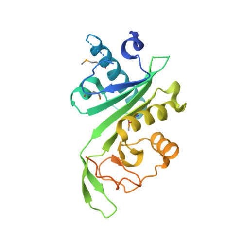
Structure of the alpha-tubulin acetyltransferase, alpha TAT1, and implications for tubulin-specific acetylation.

Friedmann, D.R.Aguilar, A.Fan, J.Nachury, M.V.Marmorstein, R.

(2012) Proc Natl Acad Sci U S A 109: 19655-19660

  • DOI: https://doi.org/10.1073/pnas.1209357109
  • Primary Citation of Related Structures:  
    4GS4

  • PubMed Abstract: 

    Protein acetylation is an important posttranslational modification with the recent identification of new substrates and enzymes, new links to disease, and modulators of protein acetylation for therapy. α-Tubulin acetyltransferase (αTAT1) is the major α-tubulin lysine-40 (K40) acetyltransferase in mammals, nematodes, and protozoa, and its activity plays a conserved role in several microtubule-based processes. Here, we present the X-ray crystal structure of the human αTAT1/acetyl-CoA complex. Together with structure-based mutagenesis, enzymatic analysis, and functional studies in cells, we elucidate the catalytic mechanism and mode of tubulin-specific acetylation. We find that αTAT1 has an overall fold similar to the Gcn5 histone acetyltransferase but contains a relatively wide substrate binding groove and unique structural elements that play important roles in α-tubulin-specific acetylation. Conserved aspartic acid and cysteine residues play important catalytic roles through a ternary complex mechanism. αTAT1 mutations have analogous effects on tubulin acetylation in vitro and in cells, demonstrating that it is the central determining factor of α-tubulin K40 acetylation levels in vivo. Together, these studies provide general insights into distinguishing features between histone and tubulin acetyltransferases, and they have specific implications for understanding the molecular basis of tubulin acetylation and for developing small molecule modulators of microtubule acetylation for therapy.


  • Organizational Affiliation

    Program in Gene Expression and Regulation, The Wistar Institute, Philadelphia, PA 19104, USA.


Macromolecules
Find similar proteins by:  (by identity cutoff)  |  3D Structure
Entity ID: 1
MoleculeChains Sequence LengthOrganismDetailsImage
Alpha-tubulin N-acetyltransferase240Homo sapiensMutation(s): 0 
Gene Names: ATAT1C6orf134MEC17Nbla00487
EC: 2.3.1.108
UniProt & NIH Common Fund Data Resources
Find proteins for Q5SQI0 (Homo sapiens)
Explore Q5SQI0 
Go to UniProtKB:  Q5SQI0
PHAROS:  Q5SQI0
GTEx:  ENSG00000137343 
Entity Groups  
Sequence Clusters30% Identity50% Identity70% Identity90% Identity95% Identity100% Identity
UniProt GroupQ5SQI0
Sequence Annotations
Expand
  • Reference Sequence
Small Molecules
Ligands 1 Unique
IDChains Name / Formula / InChI Key2D Diagram3D Interactions
ACO
Query on ACO

Download Ideal Coordinates CCD File 
B [auth A]ACETYL COENZYME *A
C23 H38 N7 O17 P3 S
ZSLZBFCDCINBPY-ZSJPKINUSA-N
Modified Residues  1 Unique
IDChains TypeFormula2D DiagramParent
MSE
Query on MSE
A
L-PEPTIDE LINKINGC5 H11 N O2 SeMET
Experimental Data & Validation

Experimental Data

  • Method: X-RAY DIFFRACTION
  • Resolution: 2.11 Å
  • R-Value Free: 0.253 
  • R-Value Work: 0.227 
  • R-Value Observed: 0.229 
  • Space Group: C 2 2 2
Unit Cell:
Length ( Å )Angle ( ˚ )
a = 97.931α = 90
b = 130.668β = 90
c = 37.423γ = 90
Software Package:
Software NamePurpose
HKL-2000data collection
SOLVEphasing
PHENIXrefinement
HKL-2000data reduction
HKL-2000data scaling

Structure Validation

View Full Validation Report


Ligand Structure Quality Assessment 


Entry History 

Deposition Data

Revision History  (Full details and data files)

  • Version 1.0: 2012-10-17
    Type: Initial release
  • Version 1.1: 2013-08-28
    Changes: Database references