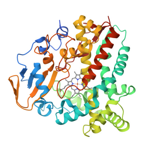
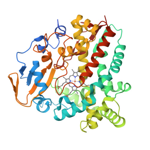
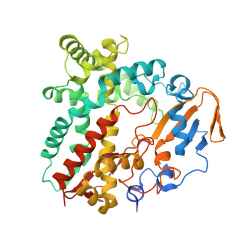
4GGV

Crystal Structure of HmtT Involved in Himastatin Biosynthesis


Experimental Data Snapshot

  • Method: X-RAY DIFFRACTION
  • Resolution: 2.33 Å
  • R-Value Free: 0.257 
  • R-Value Work: 0.208 
  • R-Value Observed: 0.210 

Starting Model: experimental
View more details

wwPDB Validation   3D Report Full Report


Ligand Structure Quality Assessment 


This is version 1.1 of the entry. See complete history


Literature

Structural analysis of HmtT and HmtN involved in the tailoring steps of himastatin biosynthesis

Zhang, H.Chen, J.Wang, H.Xie, Y.Ju, J.Yan, Y.Zhang, H.

(2013) FEBS Lett 587: 1675-1680

  • DOI: https://doi.org/10.1016/j.febslet.2013.04.013
  • Primary Citation of Related Structures:  
    4GGV

  • PubMed Abstract: 

    Himastatin is a novel antibiotic featuring a bicyclohexadepsipeptide structure. On the himastatin biosynthesis pathway, three cytochrome P450s (HmtT, HmtN, HmtS) are responsible for the post-tailoring of the cyclohexadepsipeptide backbone. Here we report the crystal structures of HmtT and HmtN. The overall structures of these two proteins are homologous to other cytochrome P450s. However, the exceptionally long F-G loop in HmtT has a highly unusual conformation and extends deep into the active site. As a result, the F/G helices of HmtT are both kinked. In contrast, the F/G helices of HmtN are straight. Also, the F/G helices in HmtT and HmtN take distinctive orientations, which may be a contributing factor for the substrate specificity of these two enzymes.


  • Organizational Affiliation

    Key Laboratory of Molecular Biophysics, Ministry of Education, Wuhan 430074, Hubei, China.


Macromolecules
Find similar proteins by:  (by identity cutoff)  |  3D Structure
Entity ID: 1
MoleculeChains Sequence LengthOrganismDetailsImage
Cytochrome P450 superfamily protein418Streptomyces himastatinicus ATCC 53653Mutation(s): 0 
Gene Names: hmtTSSOG_07642
EC: 1.14.13
UniProt
Find proteins for D9WMR2 (Streptomyces himastatinicus ATCC 53653)
Explore D9WMR2 
Go to UniProtKB:  D9WMR2
Entity Groups  
Sequence Clusters30% Identity50% Identity70% Identity90% Identity95% Identity100% Identity
UniProt GroupD9WMR2
Sequence Annotations
Expand
  • Reference Sequence
Small Molecules
Ligands 1 Unique
IDChains Name / Formula / InChI Key2D Diagram3D Interactions
HEM
Query on HEM

Download Ideal Coordinates CCD File 
B [auth A]PROTOPORPHYRIN IX CONTAINING FE
C34 H32 Fe N4 O4
KABFMIBPWCXCRK-RGGAHWMASA-L
Experimental Data & Validation

Experimental Data

  • Method: X-RAY DIFFRACTION
  • Resolution: 2.33 Å
  • R-Value Free: 0.257 
  • R-Value Work: 0.208 
  • R-Value Observed: 0.210 
  • Space Group: P 21 21 2
Unit Cell:
Length ( Å )Angle ( ˚ )
a = 64.41α = 90
b = 121.139β = 90
c = 54.731γ = 90
Software Package:
Software NamePurpose
HKL-2000data collection
SOLVEphasing
PHENIXrefinement
HKL-2000data reduction
SCALAdata scaling

Structure Validation

View Full Validation Report


Ligand Structure Quality Assessment 


Entry History 

Deposition Data

Revision History  (Full details and data files)

  • Version 1.0: 2013-07-17
    Type: Initial release
  • Version 1.1: 2023-11-08
    Changes: Data collection, Database references, Derived calculations, Refinement description