4F52

Structure of a Glomulin-RBX1-CUL1 complex


Experimental Data Snapshot

  • Method: X-RAY DIFFRACTION
  • Resolution: 3.00 Å
  • R-Value Free: 0.289 
  • R-Value Work: 0.219 
  • R-Value Observed: 0.222 

wwPDB Validation   3D Report Full Report


This is version 1.1 of the entry. See complete history


Literature

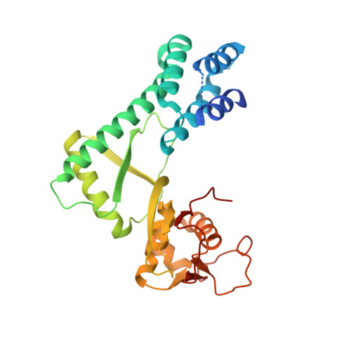
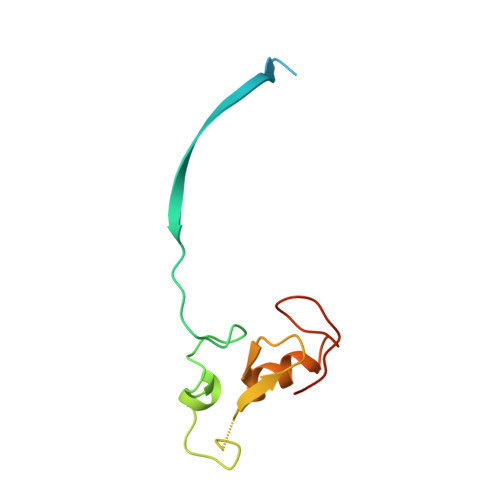
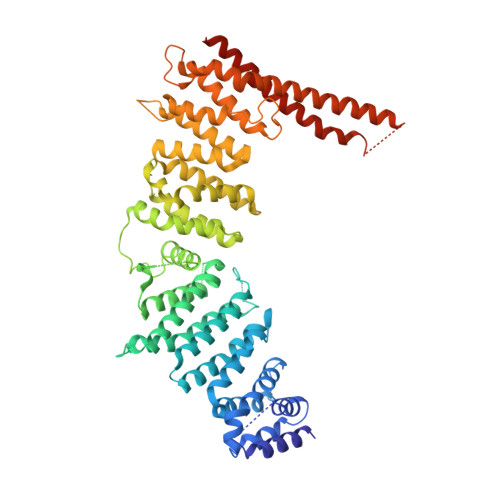
Structure of a Glomulin-RBX1-CUL1 Complex: Inhibition of a RING E3 Ligase through Masking of Its E2-Binding Surface.

Duda, D.M.Olszewski, J.L.Tron, A.E.Hammel, M.Lambert, L.J.Waddell, M.B.Mittag, T.Decaprio, J.A.Schulman, B.A.

(2012) Mol Cell 47: 371-382

  • DOI: https://doi.org/10.1016/j.molcel.2012.05.044
  • Primary Citation of Related Structures:  
    4F52

  • PubMed Abstract: 

    The approximately 300 human cullin-RING ligases (CRLs) are multisubunit E3s in which a RING protein, either RBX1 or RBX2, recruits an E2 to catalyze ubiquitination. RBX1-containing CRLs also can bind Glomulin (GLMN), which binds RBX1's RING domain, regulates the RBX1-CUL1-containing SCF(FBW7) complex, and is disrupted in the disease Glomuvenous Malformation. Here we report the crystal structure of a complex between GLMN, RBX1, and a fragment of CUL1. Structural and biochemical analyses reveal that GLMN adopts a HEAT-like repeat fold that tightly binds the E2-interacting surface of RBX1, inhibiting CRL-mediated chain formation by the E2 CDC34. The structure explains the basis for GLMN's selectivity toward RBX1 over RBX2, and how disease-associated mutations disrupt GLMN-RBX1 interactions. Our study reveals a mechanism for RING E3 ligase regulation, whereby an inhibitor blocks E2 access, and raises the possibility that other E3s are likewise controlled by cellular proteins that mask E2-binding surfaces to mediate inhibition.


  • Organizational Affiliation

    Department of Structural Biology, St. Jude Children's Research Hospital, Memphis TN 38105, USA.


Macromolecules
Find similar proteins by:  (by identity cutoff)  |  3D Structure
Entity ID: 1
MoleculeChains Sequence LengthOrganismDetailsImage
Cullin-1
A, C
282Homo sapiensMutation(s): 4 
Gene Names: CUL1
UniProt & NIH Common Fund Data Resources
Find proteins for Q13616 (Homo sapiens)
Explore Q13616 
Go to UniProtKB:  Q13616
PHAROS:  Q13616
GTEx:  ENSG00000055130 
Entity Groups  
Sequence Clusters30% Identity50% Identity70% Identity90% Identity95% Identity100% Identity
UniProt GroupQ13616
Sequence Annotations
Expand
  • Reference Sequence
Find similar proteins by:  (by identity cutoff)  |  3D Structure
Entity ID: 2
MoleculeChains Sequence LengthOrganismDetailsImage
E3 ubiquitin-protein ligase RBX1
B, D
106Homo sapiensMutation(s): 0 
Gene Names: RBX1RNF75ROC1
EC: 6.3.2
UniProt & NIH Common Fund Data Resources
Find proteins for P62877 (Homo sapiens)
Explore P62877 
Go to UniProtKB:  P62877
PHAROS:  P62877
GTEx:  ENSG00000100387 
Entity Groups  
Sequence Clusters30% Identity50% Identity70% Identity90% Identity95% Identity100% Identity
UniProt GroupP62877
Sequence Annotations
Expand
  • Reference Sequence
Find similar proteins by:  (by identity cutoff)  |  3D Structure
Entity ID: 3
MoleculeChains Sequence LengthOrganismDetailsImage
Glomulin
E, F
596Homo sapiensMutation(s): 0 
Gene Names: GLMNFAP48FAP68VMGLOM
UniProt & NIH Common Fund Data Resources
Find proteins for Q92990 (Homo sapiens)
Explore Q92990 
Go to UniProtKB:  Q92990
PHAROS:  Q92990
GTEx:  ENSG00000174842 
Entity Groups  
Sequence Clusters30% Identity50% Identity70% Identity90% Identity95% Identity100% Identity
UniProt GroupQ92990
Sequence Annotations
Expand
  • Reference Sequence
Experimental Data & Validation

Experimental Data

  • Method: X-RAY DIFFRACTION
  • Resolution: 3.00 Å
  • R-Value Free: 0.289 
  • R-Value Work: 0.219 
  • R-Value Observed: 0.222 
  • Space Group: P 1 21 1
Unit Cell:
Length ( Å )Angle ( ˚ )
a = 53.333α = 90
b = 193.932β = 98.81
c = 142.075γ = 90
Software Package:
Software NamePurpose
HKL-2000data collection
PHENIXmodel building
PHENIXrefinement
HKL-2000data reduction
HKL-2000data scaling
PHENIXphasing

Structure Validation

View Full Validation Report


Entry History 

Deposition Data

Revision History  (Full details and data files)

  • Version 1.0: 2012-09-19
    Type: Initial release
  • Version 1.1: 2024-02-28
    Changes: Data collection, Database references, Derived calculations