4ECM

2.3 Angstrom Crystal Structure of a Glucose-1-phosphate Thymidylyltransferase from Bacillus anthracis in Complex with Thymidine-5-diphospho-alpha-D-glucose and Pyrophosphate


Experimental Data Snapshot

  • Method: X-RAY DIFFRACTION
  • Resolution: 2.30 Å
  • R-Value Free: 0.186 
  • R-Value Work: 0.153 
  • R-Value Observed: 0.154 

wwPDB Validation   3D Report Full Report


Ligand Structure Quality Assessment 


This is version 1.3 of the entry. See complete history


Literature

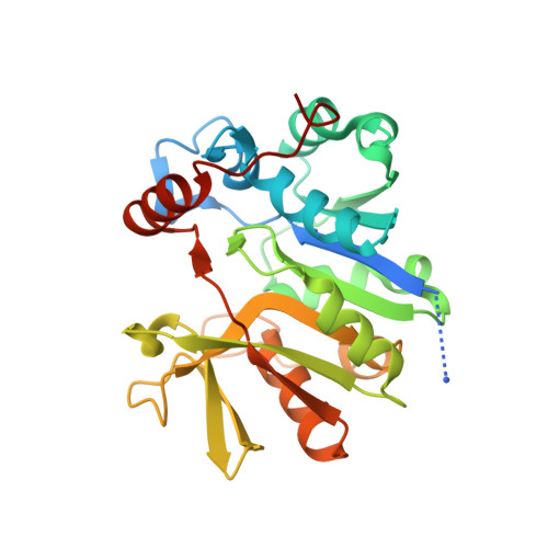
Structure of the Bacillus anthracis dTDP-L-rhamnose-biosynthetic enzyme glucose-1-phosphate thymidylyltransferase (RfbA).

Baumgartner, J.Lee, J.Halavaty, A.S.Minasov, G.Anderson, W.F.Kuhn, M.L.

(2017) Acta Crystallogr F Struct Biol Commun 73: 621-628

  • DOI: https://doi.org/10.1107/S2053230X17015357
  • Primary Citation of Related Structures:  
    4ECM

  • PubMed Abstract: 

    L-Rhamnose is a ubiquitous bacterial cell-wall component. The biosynthetic pathway for its precursor dTDP-L-rhamnose is not present in humans, which makes the enzymes of the pathway potential drug targets. In this study, the three-dimensional structure of the first protein of this pathway, glucose-1-phosphate thymidylyltransferase (RfbA), from Bacillus anthracis was determined. In other organisms this enzyme is referred to as RmlA. RfbA was co-crystallized with the products of the enzymatic reaction, dTDP-α-D-glucose and pyrophosphate, and its structure was determined at 2.3 Å resolution. This is the first reported thymidylyltransferase structure from a Gram-positive bacterium. RfbA shares overall structural characteristics with known RmlA homologs. However, RfbA exhibits a shorter sequence at its C-terminus, which results in the absence of three α-helices involved in allosteric site formation. Consequently, RfbA was observed to exhibit a quaternary structure that is unique among currently reported glucose-1-phosphate thymidylyltransferase bacterial homologs. These structural analyses suggest that RfbA may not be allosterically regulated in some organisms and is structurally distinct from other RmlA homologs.


  • Organizational Affiliation

    Department of Chemistry and Biochemistry, San Francisco State University, USA.


Macromolecules
Find similar proteins by:  (by identity cutoff)  |  3D Structure
Entity ID: 1
MoleculeChains Sequence LengthOrganismDetailsImage
Glucose-1-phosphate thymidylyltransferase269Bacillus anthracisMutation(s): 0 
Gene Names: BA1228BAS1135BA_1228GBAA_1228
EC: 2.7.7.24
UniProt
Find proteins for A0A6L8PCC3 (Bacillus anthracis)
Explore A0A6L8PCC3 
Go to UniProtKB:  A0A6L8PCC3
Entity Groups  
Sequence Clusters30% Identity50% Identity70% Identity90% Identity95% Identity100% Identity
UniProt GroupA0A6L8PCC3
Sequence Annotations
Expand
  • Reference Sequence
Small Molecules
Ligands 2 Unique
IDChains Name / Formula / InChI Key2D Diagram3D Interactions
DAU
Query on DAU

Download Ideal Coordinates CCD File 
B [auth A]2'DEOXY-THYMIDINE-5'-DIPHOSPHO-ALPHA-D-GLUCOSE
C16 H26 N2 O16 P2
YSYKRGRSMLTJNL-URARBOGNSA-N
POP
Query on POP

Download Ideal Coordinates CCD File 
C [auth A]PYROPHOSPHATE 2-
H2 O7 P2
XPPKVPWEQAFLFU-UHFFFAOYSA-L
Experimental Data & Validation

Experimental Data

Unit Cell:
Length ( Å )Angle ( ˚ )
a = 135.072α = 90
b = 135.072β = 90
c = 85.138γ = 120
Software Package:
Software NamePurpose
Blu-Icedata collection
PHASERphasing
REFMACrefinement
HKL-3000data reduction
HKL-3000data scaling

Structure Validation

View Full Validation Report


Ligand Structure Quality Assessment 


Entry History 

Revision History  (Full details and data files)

  • Version 1.0: 2012-04-04
    Type: Initial release
  • Version 1.1: 2012-05-23
    Changes: Atomic model
  • Version 1.2: 2017-11-15
    Changes: Database references, Refinement description
  • Version 1.3: 2023-09-13
    Changes: Data collection, Database references, Derived calculations, Refinement description