4EC7

Cobra NGF in complex with lipid


Experimental Data Snapshot

  • Method: X-RAY DIFFRACTION
  • Resolution: 2.60 Å
  • R-Value Free: 0.269 
  • R-Value Work: 0.228 
  • R-Value Observed: 0.230 

wwPDB Validation   3D Report Full Report


Ligand Structure Quality Assessment 


This is version 1.0 of the entry. See complete history


Literature

Structural and functional insights into lipid-bound nerve growth factors

Tong, Q.Wang, F.Zhou, H.Z.Sun, H.L.Song, H.Shu, Y.Y.Gong, Y.Zhang, W.T.Cai, T.X.Yang, F.Q.Tang, J.Jiang, T.

(2012) FASEB J 26: 3811-3821

  • DOI: https://doi.org/10.1096/fj.12-207316
  • Primary Citation of Related Structures:  
    4EAX, 4EC7

  • PubMed Abstract: 

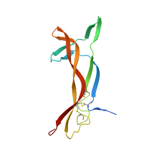
    Nerve growth factor (NGF) is a dimeric molecule that modulates the survival, proliferation, and differentiation of nervous cells and is also known to act on cells of the immune system and endocrine system. NGFs extracted from mouse submaxillary gland and cobra venom have different immunological behaviors, yet the underlying mechanism remains unclear. Here we report the crystal structure of the NGF purified from Chinese cobra Naja naja atra (cNGF), which unexpectedly reveals a 2-tailed lipid molecule that is embedded between the two protomers of the NGF homodimer. In addition, crystallographic analysis indicated that the purified mouse NGF(mNGF) is free from lipid but can bind lysophosphatidylserine (lyso-PS) in the same pocket as cNGF. Bioassays indicated that the binding of lipid molecules to cNGF and mNGF are essential for their mast cell activation activity and abates their p75(NTR) binding capacity. Taken together, these results suggest a new mechanism for the regulation of the function of NGF.


  • Organizational Affiliation

    National Key Laboratory of Biomacromolecules, Institute of Biophysics, Beijing, China.


Macromolecules
Find similar proteins by:  (by identity cutoff)  |  3D Structure
Entity ID: 1
MoleculeChains Sequence LengthOrganismDetailsImage
Venom nerve growth factor
A, B
116Naja atraMutation(s): 0 
UniProt
Find proteins for P61898 (Naja atra)
Explore P61898 
Go to UniProtKB:  P61898
Entity Groups  
Sequence Clusters30% Identity50% Identity70% Identity90% Identity95% Identity100% Identity
UniProt GroupP61898
Sequence Annotations
Expand
  • Reference Sequence
Small Molecules
Ligands 1 Unique
IDChains Name / Formula / InChI Key2D Diagram3D Interactions
L44
Query on L44

Download Ideal Coordinates CCD File 
C [auth A](2S)-1-hydroxy-3-(tetradecanoyloxy)propan-2-yl docosanoate
C39 H76 O5
DINFAYNBAABMIP-QNGWXLTQSA-N
Experimental Data & Validation

Experimental Data

  • Method: X-RAY DIFFRACTION
  • Resolution: 2.60 Å
  • R-Value Free: 0.269 
  • R-Value Work: 0.228 
  • R-Value Observed: 0.230 
  • Space Group: P 6 2 2
Unit Cell:
Length ( Å )Angle ( ˚ )
a = 118.12α = 90
b = 118.12β = 90
c = 87.35γ = 120
Software Package:
Software NamePurpose
CrystalCleardata collection
MOLREPphasing
PHENIXrefinement
CrystalCleardata reduction
CrystalCleardata scaling

Structure Validation

View Full Validation Report


Ligand Structure Quality Assessment 


Entry History 

Deposition Data

Revision History  (Full details and data files)

  • Version 1.0: 2012-09-12
    Type: Initial release