4E7V

The structure of R6 bovine insulin


Experimental Data Snapshot

  • Method: X-RAY DIFFRACTION
  • Resolution: 1.80 Å
  • R-Value Free: 0.272 
  • R-Value Work: 0.209 
  • R-Value Observed: 0.212 

wwPDB Validation   3D Report Full Report


This is version 1.3 of the entry. See complete history


Literature

The structures of T(6), T(3)R(3) and R(6) bovine insulin: combining X-ray diffraction and absorption spectroscopy.

Frankar, C.G.Knudsen, M.V.Noren, K.Nazarenko, E.Stahl, K.Harris, P.

(2012) Acta Crystallogr D Biol Crystallogr 68: 1259-1271

  • DOI: https://doi.org/10.1107/S090744491202625X
  • Primary Citation of Related Structures:  
    4E7T, 4E7U, 4E7V

  • PubMed Abstract: 

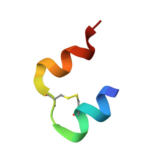
    The crystal structures of three conformations, T(6), T(3)R(3) and R(6), of bovine insulin were solved at 1.40, 1.30 and 1.80 Å resolution, respectively. All conformations crystallized in space group R3. In contrast to the T(6) and T(3)R(3) structures, different conformations of the N-terminal B-chain residue PheB1 were observed in the R(6) insulin structure, resulting in an eightfold doubling of the unit-cell volume upon cooling. The zinc coordination in each conformation was studied by X-ray absorption spectroscopy (XAS), including both EXAFS and XANES. Zinc adopts a tetrahedral coordination in all R(3) sites and an octahedral coordination in T(3) sites. The coordination distances were refined from XAS with a standard deviation of <0.01 Å. In contrast to the distances determined from the medium-resolution crystal structures, the XAS results were in good agreement with similar coordination geometries found in small molecules, as well as in other high-resolution insulin structures. As the radiation dose for XRD experiments is two orders of magnitude higher compared with that of XAS experiments, the single crystals were exposed to a higher degree of radiation damage that affected the zinc coordination in the T(3) sites in particular. Furthermore, XANES spectra for the zinc sites in T(6) and R(6) insulin were successfully calculated using finite difference methods and the bond distances and angles were optimized from a quantitative XANES analysis.


  • Organizational Affiliation

    Department of Chemistry, Technical University of Denmark, Kemitorvet B207, 2800 Kgs. Lyngby, Denmark.


Macromolecules

Find similar proteins by:  Sequence   |   3D Structure  

Entity ID: 1
MoleculeChains Sequence LengthOrganismDetailsImage
Insulin A chain21Bos taurusMutation(s): 0 
UniProt
Find proteins for P01317 (Bos taurus)
Explore P01317 
Go to UniProtKB:  P01317
Entity Groups  
Sequence Clusters30% Identity50% Identity70% Identity90% Identity95% Identity100% Identity
UniProt GroupP01317
Sequence Annotations
Expand
  • Reference Sequence
Find similar proteins by:  (by identity cutoff)  |  3D Structure
Entity ID: 2
MoleculeChains Sequence LengthOrganismDetailsImage
Insulin B chain30Bos taurusMutation(s): 0 
UniProt
Find proteins for P01317 (Bos taurus)
Explore P01317 
Go to UniProtKB:  P01317
Entity Groups  
Sequence Clusters30% Identity50% Identity70% Identity90% Identity95% Identity100% Identity
UniProt GroupP01317
Sequence Annotations
Expand
  • Reference Sequence
Small Molecules
Ligands 3 Unique
IDChains Name / Formula / InChI Key2D Diagram3D Interactions
CRS
Query on CRS

Download Ideal Coordinates CCD File 
AB [auth Y]
DB [auth 1]
GA [auth A]
GB [auth 3]
IA [auth C]
AB [auth Y],
DB [auth 1],
GA [auth A],
GB [auth 3],
IA [auth C],
JB [auth 5],
LA [auth E],
NA [auth G],
OA [auth I],
PA [auth K],
QA [auth M],
TA [auth O],
WA [auth Q],
XA [auth S],
YA [auth U],
ZA [auth W]
M-CRESOL
C7 H8 O
RLSSMJSEOOYNOY-UHFFFAOYSA-N
ZN
Query on ZN

Download Ideal Coordinates CCD File 
BB [auth Z]
EB [auth 2]
HA [auth B]
HB [auth 4]
JA [auth D]
BB [auth Z],
EB [auth 2],
HA [auth B],
HB [auth 4],
JA [auth D],
KB [auth 6],
RA [auth N],
UA [auth P]
ZINC ION
Zn
PTFCDOFLOPIGGS-UHFFFAOYSA-N
CL
Query on CL

Download Ideal Coordinates CCD File 
CB [auth Z]
FB [auth 2]
IB [auth 4]
KA [auth D]
LB [auth 6]
CB [auth Z],
FB [auth 2],
IB [auth 4],
KA [auth D],
LB [auth 6],
MA [auth F],
SA [auth N],
VA [auth P]
CHLORIDE ION
Cl
VEXZGXHMUGYJMC-UHFFFAOYSA-M
Experimental Data & Validation

Experimental Data

  • Method: X-RAY DIFFRACTION
  • Resolution: 1.80 Å
  • R-Value Free: 0.272 
  • R-Value Work: 0.209 
  • R-Value Observed: 0.212 
  • Space Group: H 3
Unit Cell:
Length ( Å )Angle ( ˚ )
a = 156.24α = 90
b = 156.24β = 90
c = 78.88γ = 120
Software Package:
Software NamePurpose
MAR345dtbdata collection
MOLREPphasing
PHENIXrefinement
XDSdata reduction
XSCALEdata scaling

Structure Validation

View Full Validation Report


Entry History 

Deposition Data

Revision History  (Full details and data files)

  • Version 1.0: 2012-04-04
    Type: Initial release
  • Version 1.1: 2012-10-03
    Changes: Database references
  • Version 1.2: 2018-03-07
    Changes: Data collection
  • Version 1.3: 2023-09-13
    Changes: Data collection, Database references, Derived calculations, Refinement description