4DOY

Crystal structure of Dibenzothiophene desulfurization enzyme C


Experimental Data Snapshot

  • Method: X-RAY DIFFRACTION
  • Resolution: 1.79 Å
  • R-Value Free: 0.194 
  • R-Value Work: 0.168 
  • R-Value Observed: 0.170 

wwPDB Validation   3D Report Full Report


This is version 1.3 of the entry. See complete history


Literature

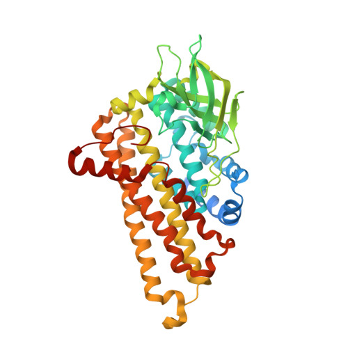
Crystal structure of DszC from Rhodococcus sp. XP at 1.79 angstrom

Liu, S.Zhang, C.Su, T.Wei, T.Zhu, D.Wang, K.Huang, Y.Dong, Y.Yin, K.Xu, S.Xu, P.Gu, L.

(2014) Proteins 82: 1708-1720

  • DOI: https://doi.org/10.1002/prot.24525
  • Primary Citation of Related Structures:  
    4DOY

  • PubMed Abstract: 

    The dibenzothiophene (DBT) monooxygenase DszC, which is the key initiating enzyme in "4S" metabolic pathway, catalyzes sequential sulphoxidation reaction of DBT to DBT sulfoxide (DBTO), then DBT sulfone (DBTO2). Here, we report the crystal structure of DszC from Rhodococcus sp. XP at 1.79 Å. Intriguingly, two distinct conformations occur in the flexible lid loops adjacent to the active site (residue 280-295, between α9 and α10). They are named "open"' and "closed" state respectively, and might show the status of the free and ligand-bound DszC. The molecular docking results suggest that the reduced FMN reacts with an oxygen molecule at C4a position of the isoalloxazine ring, producing the C4a-(hydro)peroxyflavin intermediate which is stabilized by H391 and S163. H391 may contribute to the formation of the C4a-(hydro)peroxyflavin by acting as a proton donor to the proximal peroxy oxygen, and it might also be involved in the protonation process of the C4a-(hydro)xyflavin. Site-directed mutagenesis study shows that mutations in the residues involved either in catalysis or in flavin or substrate-binding result in a complete loss of enzyme activity, suggesting that the accurate positions of flavin and substrate are crucial for the enzyme activity.


  • Organizational Affiliation

    State Key Laboratory of Microbial Technology, School of Life Sciences, Shandong University, Jinan, 250100, China.


Macromolecules
Find similar proteins by:  (by identity cutoff)  |  3D Structure
Entity ID: 1
MoleculeChains Sequence LengthOrganismDetailsImage
Dibenzothiophene desulfurization enzyme C
A, B, C, D, E
A, B, C, D, E, F, G, H
437Rhodococcus erythropolis XPMutation(s): 0 
Gene Names: dszC
EC: 1.14.14.1
UniProt
Find proteins for Q6WNP1 (Rhodococcus erythropolis (strain XP))
Explore Q6WNP1 
Go to UniProtKB:  Q6WNP1
Entity Groups  
Sequence Clusters30% Identity50% Identity70% Identity90% Identity95% Identity100% Identity
UniProt GroupQ6WNP1
Sequence Annotations
Expand
  • Reference Sequence
Small Molecules
Ligands 1 Unique
IDChains Name / Formula / InChI Key2D Diagram3D Interactions
GOL
Query on GOL

Download Ideal Coordinates CCD File 
I [auth A]
J [auth B]
K [auth B]
L [auth C]
M [auth D]
I [auth A],
J [auth B],
K [auth B],
L [auth C],
M [auth D],
N [auth D],
O [auth E],
P [auth E],
Q [auth F],
R [auth G],
S [auth G],
T [auth H],
U [auth H]
GLYCEROL
C3 H8 O3
PEDCQBHIVMGVHV-UHFFFAOYSA-N
Experimental Data & Validation

Experimental Data

  • Method: X-RAY DIFFRACTION
  • Resolution: 1.79 Å
  • R-Value Free: 0.194 
  • R-Value Work: 0.168 
  • R-Value Observed: 0.170 
  • Space Group: P 1
Unit Cell:
Length ( Å )Angle ( ˚ )
a = 95.519α = 98.46
b = 98.824β = 106.99
c = 111.465γ = 107.07
Software Package:
Software NamePurpose
HKL-2000data collection
SOLVEphasing
PHENIXrefinement
HKL-2000data reduction
HKL-2000data scaling

Structure Validation

View Full Validation Report


Entry History 

Deposition Data

Revision History  (Full details and data files)

  • Version 1.0: 2013-02-13
    Type: Initial release
  • Version 1.1: 2014-03-12
    Changes: Database references
  • Version 1.2: 2014-12-24
    Changes: Database references
  • Version 1.3: 2024-03-20
    Changes: Data collection, Database references, Derived calculations