4CAT

THREE-DIMENSIONAL STRUCTURE OF CATALASE FROM PENICILLIUM VITALE AT 2.0 ANGSTROMS RESOLUTION


Experimental Data Snapshot

  • Method: X-RAY DIFFRACTION
  • Resolution: 3.00 Å

wwPDB Validation   3D Report Full Report


This is version 2.0 of the entry. See complete history


Literature

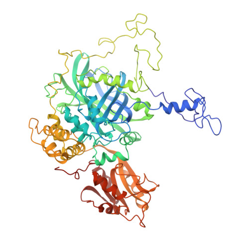
Three-dimensional structure of catalase from Penicillium vitale at 2.0 A resolution.

Vainshtein, B.K.Melik-Adamyan, W.R.Barynin, V.V.Vagin, A.A.Grebenko, A.I.Borisov, V.V.Bartels, K.S.Fita, I.Rossmann, M.G.

(1986) J Mol Biol 188: 49-61

  • DOI: https://doi.org/10.1016/0022-2836(86)90479-1
  • Primary Citation of Related Structures:  
    4CAT

  • PubMed Abstract: 

    The three-dimensional structure analysis of crystalline fungal catalase from Penicillium vitale has been extended to 2.0 A resolution. The crystals belong to space group P3(1)21, with the unit cell parameters of a = b = 144.4 A and c = 133.8 A. The asymmetric unit contains half a tetrameric molecule of 222 symmetry. Each subunit is a single polypeptide chain of approximately 670 amino acid residues and binds one heme group. The amino acid sequence has been tentatively determined by computer graphics model building (using the FRODO system) and comparison with the known sequence of beef liver catalase. The atomic model has been refined by the Hendrickson & Konnert (1981) restrained least-squares program against 68,000 reflections between 5 A and 2 A resolution. The final R-factor is 0.31 after 24 refinement cycles. The secondary and tertiary structure of the catalase has been analyzed.


Macromolecules
Find similar proteins by:  (by identity cutoff)  |  3D Structure
Entity ID: 1
MoleculeChains Sequence LengthOrganismDetailsImage
CATALASE
A, B
659Penicillium janthinellumMutation(s): 0 
EC: 1.11.1.6
Sequence Annotations
Expand
  • Reference Sequence
Small Molecules
Ligands 1 Unique
IDChains Name / Formula / InChI Key2D Diagram3D Interactions
HEM
Query on HEM

Download Ideal Coordinates CCD File 
C [auth A],
D [auth B]
PROTOPORPHYRIN IX CONTAINING FE
C34 H32 Fe N4 O4
KABFMIBPWCXCRK-RGGAHWMASA-L
Experimental Data & Validation

Experimental Data

  • Method: X-RAY DIFFRACTION
  • Resolution: 3.00 Å
  • Space Group: P 31 2 1
Unit Cell:
Length ( Å )Angle ( ˚ )
a = 144.4α = 90
b = 144.4β = 90
c = 133.8γ = 120

Structure Validation

View Full Validation Report


Entry History 

Deposition Data

Revision History  (Full details and data files)

  • Version 1.0: 1983-09-06
    Type: Initial release
  • Version 1.1: 2008-03-25
    Changes: Version format compliance
  • Version 1.2: 2011-07-13
    Changes: Advisory, Version format compliance
  • Version 1.3: 2023-05-31
    Changes: Database references, Other
  • Version 2.0: 2023-09-27
    Type: Remediation
    Changes: Advisory, Atomic model, Data collection, Derived calculations, Other, Refinement description