4B6C

Structure of the M. smegmatis GyrB ATPase domain in complex with an aminopyrazinamide


Experimental Data Snapshot

  • Method: X-RAY DIFFRACTION
  • Resolution: 2.20 Å
  • R-Value Free: 0.254 
  • R-Value Work: 0.199 
  • R-Value Observed: 0.202 

wwPDB Validation   3D Report Full Report


Ligand Structure Quality Assessment 


This is version 1.3 of the entry. See complete history


Literature

Aminopyrazinamides: novel and specific GyrB inhibitors that kill replicating and nonreplicating Mycobacterium tuberculosis.

Shirude, P.S.Madhavapeddi, P.Tucker, J.A.Murugan, K.Patil, V.Basavarajappa, H.Raichurkar, A.V.Humnabadkar, V.Hussein, S.Sharma, S.Ramya, V.K.Narayan, C.B.Balganesh, T.S.Sambandamurthy, V.K.

(2013) ACS Chem Biol 8: 519-523

  • DOI: https://doi.org/10.1021/cb300510w
  • Primary Citation of Related Structures:  
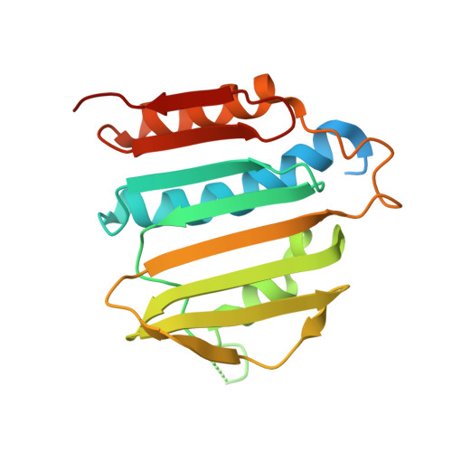
    4B6C

  • PubMed Abstract: 

    Aminopyrazinamides originated from a high throughput screen targeting the Mycobacterium smegmatis (Msm) GyrB ATPase. This series displays chemical tractability, robust structure-activity relationship, and potent antitubercular activity. The crystal structure of Msm GyrB in complex with one of the aminopyrazinamides revealed promising attributes of specificity against other broad spectrum pathogens and selectivity against eukaryotic kinases due to novel interactions at hydrophobic pocket, unlike other known GyrB inhibitors. The aminopyrazinamides display excellent mycobacterial kill under in vitro, intracellular, and hypoxic conditions.


  • Organizational Affiliation

    Department of Medicinal Chemistry, AstraZeneca India Pvt. Ltd., Bellary Road, Hebbal, Bangalore 560024, India. pravin.shirude@astrazeneca.com


Macromolecules
Find similar proteins by:  (by identity cutoff)  |  3D Structure
Entity ID: 1
MoleculeChains Sequence LengthOrganismDetailsImage
DNA gyrase subunit B,DNA gyrase subunit B,DNA gyrase subunit B
A, B
196Mycolicibacterium smegmatisMutation(s): 2 
Gene Names: gyrB
EC: 5.99.1.3
UniProt
Find proteins for P0C559 (Mycolicibacterium smegmatis)
Explore P0C559 
Go to UniProtKB:  P0C559
Entity Groups  
Sequence Clusters30% Identity50% Identity70% Identity90% Identity95% Identity100% Identity
UniProt GroupP0C559
Sequence Annotations
Expand
  • Reference Sequence
Small Molecules
Binding Affinity Annotations 
IDSourceBinding Affinity
B5U Binding MOAD:  4B6C IC50: 690 (nM) from 1 assay(s)
PDBBind:  4B6C IC50: 690 (nM) from 1 assay(s)
Experimental Data & Validation

Experimental Data

  • Method: X-RAY DIFFRACTION
  • Resolution: 2.20 Å
  • R-Value Free: 0.254 
  • R-Value Work: 0.199 
  • R-Value Observed: 0.202 
  • Space Group: C 2 2 21
Unit Cell:
Length ( Å )Angle ( ˚ )
a = 43.455α = 90
b = 82.251β = 90
c = 191.941γ = 90
Software Package:
Software NamePurpose
BUSTERrefinement
d*TREKdata reduction
d*TREKdata scaling
AMoREphasing

Structure Validation

View Full Validation Report


Ligand Structure Quality Assessment 


Entry History 

Deposition Data

Revision History  (Full details and data files)

  • Version 1.0: 2013-01-23
    Type: Initial release
  • Version 1.1: 2013-04-03
    Changes: Database references, Structure summary
  • Version 1.2: 2018-04-04
    Changes: Data collection
  • Version 1.3: 2018-11-28
    Changes: Data collection, Database references, Source and taxonomy, Structure summary