4AL9

Crystal structure of the lectin PA-IL from Pseudomonas aeruginoas in complex with melibiose


Experimental Data Snapshot

  • Method: X-RAY DIFFRACTION
  • Resolution: 1.75 Å
  • R-Value Free: 0.211 
  • R-Value Work: 0.167 
  • R-Value Observed: 0.169 

wwPDB Validation   3D Report Full Report


Ligand Structure Quality Assessment 


This is version 2.1 of the entry. See complete history


Literature

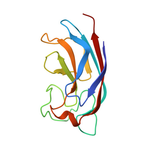
Secondary Sugar Binding Site Identified for Leca Lectin from Pseudomonas Aeruginosa.

Blanchard, B.Imberty, A.Varrot, A.

(2014) Proteins 82: 1060

  • DOI: https://doi.org/10.1002/prot.24430
  • Primary Citation of Related Structures:  
    4AL9

  • PubMed Abstract: 

    The galactose-specific lectin LecA from Pseudomonas aeruginosa is a target for the development of new anti-infectious compounds. Sugar based molecules with anti-adhesive properties present great potential in the fight against bacterial infection and biofilm formation. LecA is specific for oligosaccharides with terminal α-galactoside residues and displays strong affinity for melibiose (αGal1-6Glc) with a Kd of 38.8 µM. The crystal structure of LecA/melibiose complex shows classical calcium-bridged binding of αGal in the primary binding site but also revealed a secondary sugar binding site with glucose bound. This sugar binding site is in close proximity to the galactose binding one, is independent of calcium and mainly involves interactions with a symmetry-related protein. This discovery would help to the design of new potent inhibitors targeting both binding sites.


  • Organizational Affiliation

    CERMAV-CNRS, Université de Grenoble and Member of ICMG, BP 53, 38041 Grenoble Cedex 9, France.


Macromolecules
Find similar proteins by:  (by identity cutoff)  |  3D Structure
Entity ID: 1
MoleculeChains Sequence LengthOrganismDetailsImage
PA-I GALACTOPHILIC LECTIN
A, B, C, D, E
A, B, C, D, E, F, G, H
121Pseudomonas aeruginosa PAO1Mutation(s): 0 
UniProt
Find proteins for Q05097 (Pseudomonas aeruginosa (strain ATCC 15692 / DSM 22644 / CIP 104116 / JCM 14847 / LMG 12228 / 1C / PRS 101 / PAO1))
Explore Q05097 
Go to UniProtKB:  Q05097
Entity Groups  
Sequence Clusters30% Identity50% Identity70% Identity90% Identity95% Identity100% Identity
UniProt GroupQ05097
Sequence Annotations
Expand
  • Reference Sequence
Oligosaccharides

Help

Entity ID: 2
MoleculeChains Length2D Diagram Glycosylation3D Interactions
alpha-D-galactopyranose-(1-6)-alpha-D-glucopyranose
I, J
2N/A
Glycosylation Resources
GlyTouCan:  G54976AD
GlyCosmos:  G54976AD
GlyGen:  G54976AD
Small Molecules
Ligands 3 Unique
IDChains Name / Formula / InChI Key2D Diagram3D Interactions
GLA
Query on GLA

Download Ideal Coordinates CCD File 
AA [auth G]
L [auth A]
O [auth B]
Q [auth C]
S [auth D]
AA [auth G],
L [auth A],
O [auth B],
Q [auth C],
S [auth D],
V [auth E]
alpha-D-galactopyranose
C6 H12 O6
WQZGKKKJIJFFOK-PHYPRBDBSA-N
EDO
Query on EDO

Download Ideal Coordinates CCD File 
BA [auth H],
M [auth A],
T [auth D],
W [auth F],
Y [auth F]
1,2-ETHANEDIOL
C2 H6 O2
LYCAIKOWRPUZTN-UHFFFAOYSA-N
CA
Query on CA

Download Ideal Coordinates CCD File 
CA [auth H]
K [auth A]
N [auth B]
P [auth C]
R [auth D]
CA [auth H],
K [auth A],
N [auth B],
P [auth C],
R [auth D],
U [auth E],
X [auth F],
Z [auth G]
CALCIUM ION
Ca
BHPQYMZQTOCNFJ-UHFFFAOYSA-N
Biologically Interesting Molecules (External Reference) 1 Unique
Experimental Data & Validation

Experimental Data

  • Method: X-RAY DIFFRACTION
  • Resolution: 1.75 Å
  • R-Value Free: 0.211 
  • R-Value Work: 0.167 
  • R-Value Observed: 0.169 
  • Space Group: P 1
Unit Cell:
Length ( Å )Angle ( ˚ )
a = 50.07α = 101.13
b = 58.13β = 92.89
c = 75.94γ = 100.98
Software Package:
Software NamePurpose
REFMACrefinement
iMOSFLMdata reduction
SCALAdata scaling
PHASERphasing

Structure Validation

View Full Validation Report


Ligand Structure Quality Assessment 


Entry History 

Deposition Data

Revision History  (Full details and data files)

  • Version 1.0: 2013-03-20
    Type: Initial release
  • Version 1.1: 2013-10-30
    Changes: Database references
  • Version 1.2: 2014-05-28
    Changes: Database references
  • Version 1.3: 2015-09-16
    Changes: Structure summary
  • Version 2.0: 2020-07-29
    Type: Remediation
    Reason: Carbohydrate remediation
    Changes: Atomic model, Data collection, Derived calculations, Other, Structure summary
  • Version 2.1: 2023-12-20
    Changes: Data collection, Database references, Refinement description, Structure summary