4A8E

The structure of a dimeric Xer recombinase from archaea


Experimental Data Snapshot

  • Method: X-RAY DIFFRACTION
  • Resolution: 2.99 Å
  • R-Value Free: 0.293 
  • R-Value Work: 0.207 
  • R-Value Observed: 0.211 

wwPDB Validation   3D Report Full Report


This is version 1.2 of the entry. See complete history


Literature

The Carboxy-Terminal Alpha N Helix of the Archaeal Xera Tyrosine Recombinase is a Molecular Switch to Control Site-Specific Recombination.

Serre, M.C.El Arnaout, T.Brooks, M.A.Durand, D.Lisboa, J.Lazar, N.Raynal, B.Van Tilbeurgh, H.Quevillon-Cheruel, S.

(2013) PLoS One 8: 63010

  • DOI: https://doi.org/10.1371/journal.pone.0063010
  • Primary Citation of Related Structures:  
    4A8E

  • PubMed Abstract: 

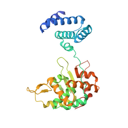
    Tyrosine recombinases are conserved in the three kingdoms of life. Here we present the first crystal structure of a full-length archaeal tyrosine recombinase, XerA from Pyrococcus abyssi, at 3.0 Å resolution. In the absence of DNA substrate XerA crystallizes as a dimer where each monomer displays a tertiary structure similar to that of DNA-bound Tyr-recombinases. Active sites are assembled in the absence of dif except for the catalytic Tyr, which is extruded and located equidistant from each active site within the dimer. Using XerA active site mutants we demonstrate that XerA follows the classical cis-cleavage reaction, suggesting rearrangements of the C-terminal domain upon DNA binding. Surprisingly, XerA C-terminal αN helices dock in cis in a groove that, in bacterial tyrosine recombinases, accommodates in trans αN helices of neighbour monomers in the Holliday junction intermediates. Deletion of the XerA C-terminal αN helix does not impair cleavage of suicide substrates but prevents recombination catalysis. We propose that the enzymatic cycle of XerA involves the switch of the αN helix from cis to trans packing, leading to (i) repositioning of the catalytic Tyr in the active site in cis and (ii) dimer stabilisation via αN contacts in trans between monomers.


  • Organizational Affiliation

    Institut de Génétique et Microbiologie, Université Paris-Sud, Orsay, France. marie-claude.serre@igmors.u-psud.fr


Macromolecules
Find similar proteins by:  (by identity cutoff)  |  3D Structure
Entity ID: 1
MoleculeChains Sequence LengthOrganismDetailsImage
PROBABLE TYROSINE RECOMBINASE XERC-LIKE292Pyrococcus abyssiMutation(s): 0 
UniProt
Find proteins for Q9V1P5 (Pyrococcus abyssi (strain GE5 / Orsay))
Explore Q9V1P5 
Go to UniProtKB:  Q9V1P5
Entity Groups  
Sequence Clusters30% Identity50% Identity70% Identity90% Identity95% Identity100% Identity
UniProt GroupQ9V1P5
Sequence Annotations
Expand
  • Reference Sequence
Small Molecules
Ligands 3 Unique
IDChains Name / Formula / InChI Key2D Diagram3D Interactions
SO4
Query on SO4

Download Ideal Coordinates CCD File 
B [auth A]
C [auth A]
D [auth A]
E [auth A]
F [auth A]
B [auth A],
C [auth A],
D [auth A],
E [auth A],
F [auth A],
G [auth A],
H [auth A]
SULFATE ION
O4 S
QAOWNCQODCNURD-UHFFFAOYSA-L
EDO
Query on EDO

Download Ideal Coordinates CCD File 
J [auth A]1,2-ETHANEDIOL
C2 H6 O2
LYCAIKOWRPUZTN-UHFFFAOYSA-N
CL
Query on CL

Download Ideal Coordinates CCD File 
I [auth A]CHLORIDE ION
Cl
VEXZGXHMUGYJMC-UHFFFAOYSA-M
Experimental Data & Validation

Experimental Data

  • Method: X-RAY DIFFRACTION
  • Resolution: 2.99 Å
  • R-Value Free: 0.293 
  • R-Value Work: 0.207 
  • R-Value Observed: 0.211 
  • Space Group: C 2 2 2
Unit Cell:
Length ( Å )Angle ( ˚ )
a = 92.67α = 90
b = 157.29β = 90
c = 45.55γ = 90
Software Package:
Software NamePurpose
REFMACrefinement
XDSdata reduction
XSCALEdata scaling
PHASERphasing

Structure Validation

View Full Validation Report


Entry History 

Deposition Data

Revision History  (Full details and data files)

  • Version 1.0: 2012-12-05
    Type: Initial release
  • Version 1.1: 2014-01-15
    Changes: Database references
  • Version 1.2: 2023-12-20
    Changes: Data collection, Database references, Derived calculations, Other, Refinement description