4ZI9

Structure of mouse clustered PcdhgA1 EC1-3


Experimental Data Snapshot

  • Method: X-RAY DIFFRACTION
  • Resolution: 1.70 Å
  • R-Value Free: 0.266 
  • R-Value Work: 0.230 
  • R-Value Observed: 0.231 

wwPDB Validation   3D Report Full Report


This is version 1.5 of the entry. See complete history


Literature

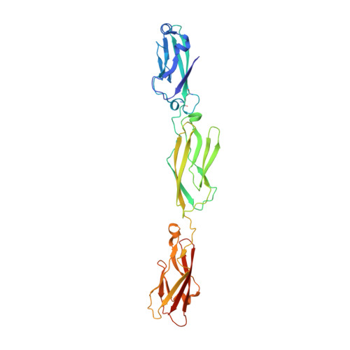
Structure and Sequence Analyses of Clustered Protocadherins Reveal Antiparallel Interactions that Mediate Homophilic Specificity.

Nicoludis, J.M.Lau, S.Y.Scharfe, C.P.Marks, D.S.Weihofen, W.A.Gaudet, R.

(2015) Structure 23: 2087-2098

  • DOI: https://doi.org/10.1016/j.str.2015.09.005
  • Primary Citation of Related Structures:  
    4ZI8, 4ZI9

  • PubMed Abstract: 

    Clustered protocadherin (Pcdh) proteins mediate dendritic self-avoidance in neurons via specific homophilic interactions in their extracellular cadherin (EC) domains. We determined crystal structures of EC1-EC3, containing the homophilic specificity-determining region, of two mouse clustered Pcdh isoforms (PcdhγA1 and PcdhγC3) to investigate the nature of the homophilic interaction. Within the crystal lattices, we observe antiparallel interfaces consistent with a role in trans cell-cell contact. Antiparallel dimerization is supported by evolutionary correlations. Two interfaces, located primarily on EC2-EC3, involve distinctive clustered Pcdh structure and sequence motifs, lack predicted glycosylation sites, and contain residues highly conserved in orthologs but not paralogs, pointing toward their biological significance as homophilic interaction interfaces. These two interfaces are similar yet distinct, reflecting a possible difference in interaction architecture between clustered Pcdh subfamilies. These structures initiate a molecular understanding of clustered Pcdh assemblies that are required to produce functional neuronal networks.


  • Organizational Affiliation

    Department of Chemistry and Chemical Biology, Harvard University, 12 Oxford Street, Cambridge, MA 02138, USA.


Macromolecules
Find similar proteins by:  (by identity cutoff)  |  3D Structure
Entity ID: 1
MoleculeChains Sequence LengthOrganismDetailsImage
MCG133388, isoform CRA_t
A, B
318Mus musculusMutation(s): 0 
Gene Names: Pcdhga1mCG_133388
UniProt
Find proteins for Q91XZ0 (Mus musculus)
Explore Q91XZ0 
Go to UniProtKB:  Q91XZ0
Entity Groups  
Sequence Clusters30% Identity50% Identity70% Identity90% Identity95% Identity100% Identity
UniProt GroupQ91XZ0
Sequence Annotations
Expand
  • Reference Sequence
Small Molecules
Ligands 1 Unique
IDChains Name / Formula / InChI Key2D Diagram3D Interactions
CA
Query on CA

Download Ideal Coordinates CCD File 
C [auth A]
D [auth A]
E [auth A]
F [auth A]
G [auth A]
C [auth A],
D [auth A],
E [auth A],
F [auth A],
G [auth A],
H [auth A],
I [auth B],
J [auth B],
K [auth B],
L [auth B],
M [auth B],
N [auth B]
CALCIUM ION
Ca
BHPQYMZQTOCNFJ-UHFFFAOYSA-N
Experimental Data & Validation

Experimental Data

Unit Cell:
Length ( Å )Angle ( ˚ )
a = 67.751α = 90
b = 63.687β = 99.69
c = 107.486γ = 90
Software Package:
Software NamePurpose
PHENIXrefinement
HKL-2000data reduction
HKL-2000data scaling
PHASERphasing

Structure Validation

View Full Validation Report


Entry History & Funding Information

Deposition Data


Funding OrganizationLocationGrant Number
National Institutes of Health/National Institute of General Medical Sciences (NIH/NIGMS)United StatesP41 GM103403
National Institutes of Health/National Center for Research Resources (NIH/NCRR)United StatesS10 RR029205
Department of Energy (DOE, United States)United StatesDE-AC02-06CH11357

Revision History  (Full details and data files)

  • Version 1.0: 2015-10-28
    Type: Initial release
  • Version 1.1: 2015-11-04
    Changes: Database references
  • Version 1.2: 2015-11-18
    Changes: Database references
  • Version 1.3: 2017-09-13
    Changes: Author supporting evidence, Database references, Derived calculations
  • Version 1.4: 2019-12-04
    Changes: Author supporting evidence
  • Version 1.5: 2023-09-27
    Changes: Data collection, Database references, Derived calculations, Refinement description