4YXH

Crystal structure of Deer prion protein complexed with POM1 FAB


Experimental Data Snapshot

  • Method: X-RAY DIFFRACTION
  • Resolution: 2.70 Å
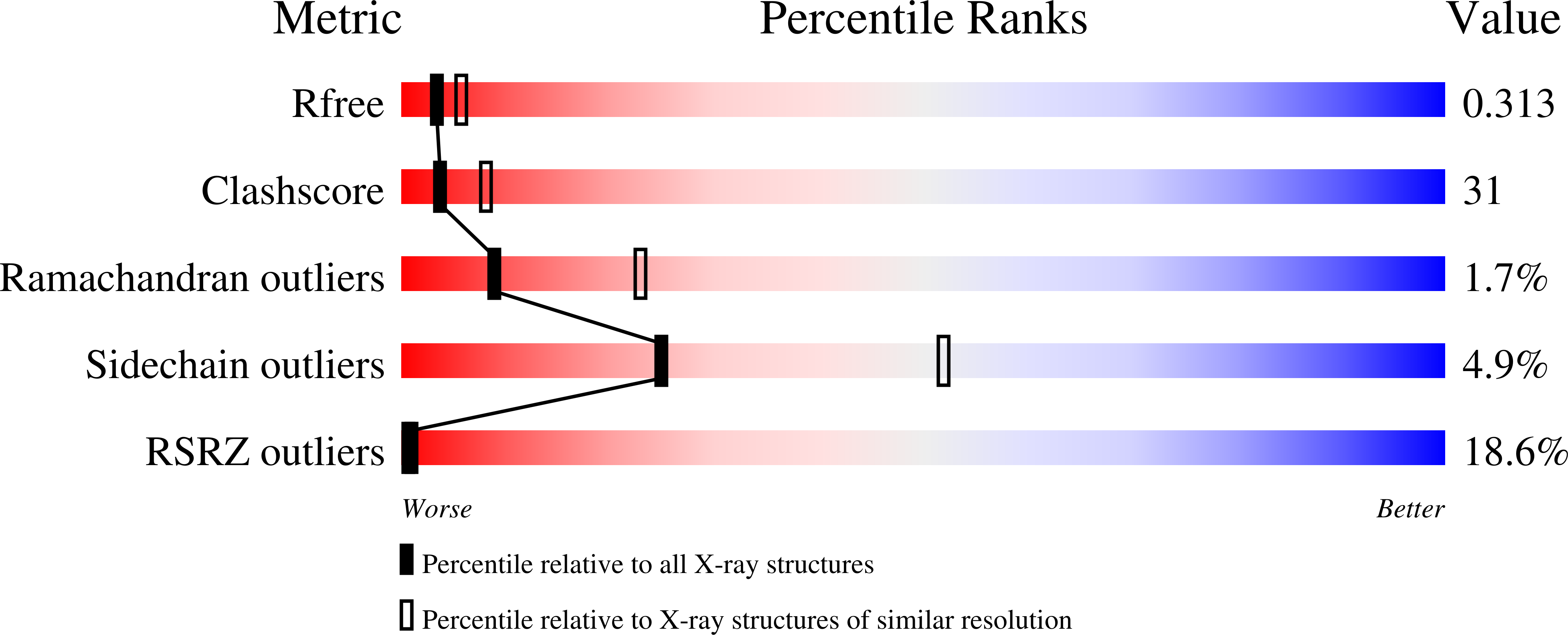
  • R-Value Free: 0.288 
  • R-Value Work: 0.242 
  • R-Value Observed: 0.245 

wwPDB Validation   3D Report Full Report


This is version 1.2 of the entry. See complete history


Literature

X-ray structural and molecular dynamical studies of the globular domains of cow, deer, elk and Syrian hamster prion proteins.

Baral, P.K.Swayampakula, M.Aguzzi, A.James, M.N.

(2015) J Struct Biol 192: 37-47

  • DOI: https://doi.org/10.1016/j.jsb.2015.08.014
  • Primary Citation of Related Structures:  
    4YX2, 4YXH, 4YXK, 4YXL

  • PubMed Abstract: 

    Misfolded prion proteins are the cause of neurodegenerative diseases that affect many mammalian species, including humans. Transmission of the prion diseases poses a considerable public-health risk as a specific prion disease such as bovine spongiform encephalopathy can be transferred to humans and other mammalian species upon contaminant exposure. The underlying mechanism of prion propagation and the species barriers that control cross species transmission has been investigated quite extensively. So far a number of prion strains have been characterized and those have been intimately linked to species-specific infectivity and other pathophysiological manifestations. These strains are encoded by a protein-only agent, and have a high degree of sequence identity across mammalian species. The molecular events that lead to strain differentiation remain elusive. In order to contribute to the understanding of strain differentiation, we have determined the crystal structures of the globular, folded domains of four prion proteins (cow, deer, elk and Syrian hamster) bound to the POM1 antibody fragment Fab. Although the overall structural folds of the mammalian prion proteins remains extremely similar, there are several local structural variations observed in the misfolding-initiator motifs. In additional molecular dynamics simulation studies on these several prion proteins reveal differences in the local fluctuations and imply that these differences have possible roles in the unfolding of the globular domains. These local variations in the structured domains perpetuate diverse patterns of prion misfolding and possibly facilitate the strain selection and adaptation.


  • Organizational Affiliation

    Department of Biochemistry, Faculty of Medicine and Dentistry, University of Alberta, Edmonton, AB T6G 2H7, Canada.


Macromolecules
Find similar proteins by:  (by identity cutoff)  |  3D Structure
Entity ID: 1
MoleculeChains Sequence LengthOrganismDetailsImage
Major prion protein135Odocoileus hemionusMutation(s): 0 
Gene Names: PRNPPRP
UniProt
Find proteins for P47852 (Odocoileus hemionus)
Explore P47852 
Go to UniProtKB:  P47852
Entity Groups  
Sequence Clusters30% Identity50% Identity70% Identity90% Identity95% Identity100% Identity
UniProt GroupP47852
Sequence Annotations
Expand
  • Reference Sequence
Find similar proteins by:  (by identity cutoff)  |  3D Structure
Entity ID: 2
MoleculeChains Sequence LengthOrganismDetailsImage
POM1 FAB HEAVY CHAINB [auth H]218Mus musculusMutation(s): 0 
Entity Groups  
Sequence Clusters30% Identity50% Identity70% Identity90% Identity95% Identity100% Identity
Sequence Annotations
Expand
  • Reference Sequence
Find similar proteins by:  (by identity cutoff)  |  3D Structure
Entity ID: 3
MoleculeChains Sequence LengthOrganismDetailsImage
POM1 FAB LIGHT CHAINC [auth L]213Mus musculusMutation(s): 0 
Entity Groups  
Sequence Clusters30% Identity50% Identity70% Identity90% Identity95% Identity100% Identity
Sequence Annotations
Expand
  • Reference Sequence
Small Molecules
Ligands 1 Unique
IDChains Name / Formula / InChI Key2D Diagram3D Interactions
NA
Query on NA

Download Ideal Coordinates CCD File 
D [auth L]SODIUM ION
Na
FKNQFGJONOIPTF-UHFFFAOYSA-N
Experimental Data & Validation

Experimental Data

  • Method: X-RAY DIFFRACTION
  • Resolution: 2.70 Å
  • R-Value Free: 0.288 
  • R-Value Work: 0.242 
  • R-Value Observed: 0.245 
  • Space Group: C 1 2 1
Unit Cell:
Length ( Å )Angle ( ˚ )
a = 84.282α = 90
b = 106.58β = 95.81
c = 75.775γ = 90
Software Package:
Software NamePurpose
PHENIXrefinement
HKL-2000data reduction
HKL-2000data scaling
MOLREPphasing

Structure Validation

View Full Validation Report


Entry History 

Deposition Data

Revision History  (Full details and data files)

  • Version 1.0: 2015-09-23
    Type: Initial release
  • Version 1.1: 2015-10-14
    Changes: Database references
  • Version 1.2: 2023-09-27
    Changes: Data collection, Database references, Derived calculations, Refinement description