4W8P

Crystal structure of RIAM TBS1 in complex with talin R7R8 domains


Experimental Data Snapshot

  • Method: X-RAY DIFFRACTION
  • Resolution: 1.50 Å
  • R-Value Free: 0.242 
  • R-Value Work: 0.223 
  • R-Value Observed: 0.224 

wwPDB Validation   3D Report Full Report


This is version 2.0 of the entry. See complete history


Literature

Structural and Mechanistic Insights into the Recruitment of Talin by RIAM in Integrin Signaling.

Chang, Y.C.Zhang, H.Franco-Barraza, J.Brennan, M.L.Patel, T.Cukierman, E.Wu, J.

(2014) Structure 22: 1810-1820

  • DOI: https://doi.org/10.1016/j.str.2014.09.020
  • Primary Citation of Related Structures:  
    4W8P

  • PubMed Abstract: 

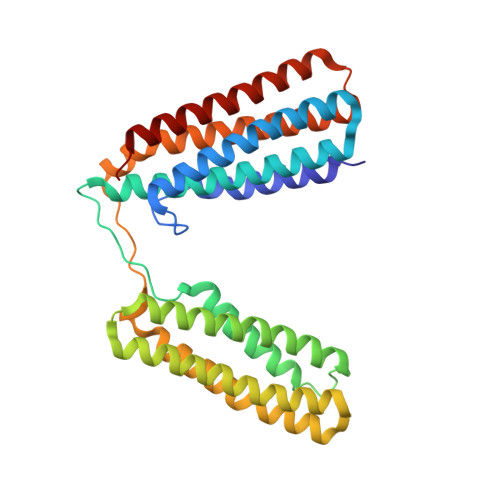
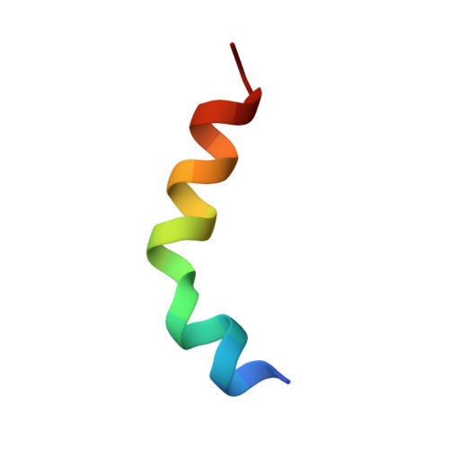
    Plasma membrane (PM)-bound GTPase Rap1 recruits the Rap1-interacting-adaptor-molecule (RIAM), which in turn recruits talin to bind and activate integrins. However, it is unclear how RIAM recruits talin and why its close homolog lamellipodin does not. Here, we report that, although RIAM possesses two talin-binding sites (TBS1 and TBS2), only TBS1 is capable of recruiting cytoplasmic talin to the PM, and the R8 domain is the strongest binding site in talin. Crystal structure of an R7R8:TBS1 complex reveals an unexpected kink in the TBS1 helix that is not shared in the homologous region of lamellipodin. This kinked helix conformation is required for the colocalization of RIAM and talin at the PM and proper activation of integrin. Our findings provide the structural and mechanistic insight into talin recruitment by RIAM that underlies integrin activation and explain the differential functions of the otherwise highly homologous RIAM and lamellipodin in integrin signaling.


  • Organizational Affiliation

    Developmental Therapeutics Program, Fox Chase Cancer Center, 333 Cottman Avenue, Philadelphia, PA 19111, USA.


Macromolecules
Find similar proteins by:  (by identity cutoff)  |  3D Structure
Entity ID: 1
MoleculeChains Sequence LengthOrganismDetailsImage
Talin-1301Mus musculusMutation(s): 0 
Gene Names: Tln1Tln
UniProt
Find proteins for P26039 (Mus musculus)
Explore P26039 
Go to UniProtKB:  P26039
Entity Groups  
Sequence Clusters30% Identity50% Identity70% Identity90% Identity95% Identity100% Identity
UniProt GroupP26039
Sequence Annotations
Expand
  • Reference Sequence

Find similar proteins by:  Sequence   |   3D Structure  

Entity ID: 2
MoleculeChains Sequence LengthOrganismDetailsImage
Amyloid beta A4 precursor protein-binding family B member 1-interacting protein21Mus musculusMutation(s): 0 
UniProt & NIH Common Fund Data Resources
Find proteins for Q8R5A3 (Mus musculus)
Explore Q8R5A3 
Go to UniProtKB:  Q8R5A3
IMPC:  MGI:1861354
Entity Groups  
Sequence Clusters30% Identity50% Identity70% Identity90% Identity95% Identity100% Identity
UniProt GroupQ8R5A3
Sequence Annotations
Expand
  • Reference Sequence
Experimental Data & Validation

Experimental Data

  • Method: X-RAY DIFFRACTION
  • Resolution: 1.50 Å
  • R-Value Free: 0.242 
  • R-Value Work: 0.223 
  • R-Value Observed: 0.224 
  • Space Group: P 21 21 2
Unit Cell:
Length ( Å )Angle ( ˚ )
a = 59.722α = 90
b = 105.884β = 90
c = 49.027γ = 90
Software Package:
Software NamePurpose
REFMACrefinement

Structure Validation

View Full Validation Report


Entry History 

Deposition Data

Revision History  (Full details and data files)

  • Version 1.0: 2014-12-03
    Type: Initial release
  • Version 1.1: 2014-12-17
    Changes: Database references
  • Version 2.0: 2023-09-27
    Changes: Atomic model, Data collection, Database references, Derived calculations, Other, Refinement description, Source and taxonomy