4PZT

Crystal structure of p300 histone acetyltransferase domain in complex with an inhibitor, Acetonyl-Coenzyme A


Experimental Data Snapshot

  • Method: X-RAY DIFFRACTION
  • Resolution: 2.80 Å
  • R-Value Free: 0.236 
  • R-Value Work: 0.157 
  • R-Value Observed: 0.161 

wwPDB Validation   3D Report Full Report


Ligand Structure Quality Assessment 


This is version 1.2 of the entry. See complete history


Literature

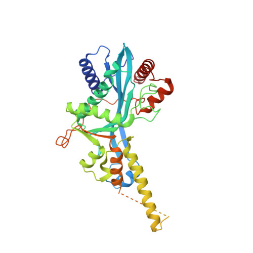
Structure of the p300 Histone Acetyltransferase Bound to Acetyl-Coenzyme A and Its Analogues.

Maksimoska, J.Segura-Pena, D.Cole, P.A.Marmorstein, R.

(2014) Biochemistry 53: 3415-3422

  • DOI: https://doi.org/10.1021/bi500380f
  • Primary Citation of Related Structures:  
    4PZR, 4PZS, 4PZT

  • PubMed Abstract: 

    The p300 and CBP transcriptional coactivator paralogs (p300/CBP) regulate a variety of different cellular pathways, in part, by acetylating histones and more than 70 non-histone protein substrates. Mutation, chromosomal translocation, or other aberrant activities of p300/CBP are linked to many different diseases, including cancer. Because of its pleiotropic biological roles and connection to disease, it is important to understand the mechanism of acetyl transfer by p300/CBP, in part so that inhibitors can be more rationally developed. Toward this goal, a structure of p300 bound to a Lys-CoA bisubstrate HAT inhibitor has been previously elucidated, and the enzyme's catalytic mechanism has been investigated. Nonetheless, many questions underlying p300/CBP structure and mechanism remain. Here, we report a structural characterization of different reaction states in the p300 activity cycle. We present the structures of p300 in complex with an acetyl-CoA substrate, a CoA product, and an acetonyl-CoA inhibitor. A comparison of these structures with the previously reported p300/Lys-CoA complex demonstrates that the conformation of the enzyme active site depends on the interaction of the enzyme with the cofactor, and is not apparently influenced by protein substrate lysine binding. The p300/CoA crystals also contain two poly(ethylene glycol) moieties bound proximal to the cofactor binding site, implicating the path of protein substrate association. The structure of the p300/acetonyl-CoA complex explains the inhibitory and tight binding properties of the acetonyl-CoA toward p300. Together, these studies provide new insights into the molecular basis of acetylation by p300 and have implications for the rational development of new small molecule p300 inhibitors.


  • Organizational Affiliation

    Program in Gene Expression and Regulation, The Wistar Institute , 3601 Spruce Street, Philadelphia, Pennsylvania 19104, United States.


Macromolecules
Find similar proteins by:  (by identity cutoff)  |  3D Structure
Entity ID: 1
MoleculeChains Sequence LengthOrganismDetailsImage
Histone acetyltransferase p300378Homo sapiensMutation(s): 1 
Gene Names: EP300P300
EC: 2.3.1.48
UniProt & NIH Common Fund Data Resources
Find proteins for Q09472 (Homo sapiens)
Explore Q09472 
Go to UniProtKB:  Q09472
PHAROS:  Q09472
GTEx:  ENSG00000100393 
Entity Groups  
Sequence Clusters30% Identity50% Identity70% Identity90% Identity95% Identity100% Identity
UniProt GroupQ09472
Sequence Annotations
Expand
  • Reference Sequence
Small Molecules
Ligands 2 Unique
IDChains Name / Formula / InChI Key2D Diagram3D Interactions
SOP
Query on SOP

Download Ideal Coordinates CCD File 
B [auth A][(2R,3S,4R,5R)-5-(6-AMINO-9H-PURIN-9-YL)-4-HYDROXY-3-(PHOSPHONOOXY)TETRAHYDROFURAN-2-YL]METHYL (3R)-3-HYDROXY-2,2-DIMETHYL-4-OXO-4-{[3-OXO-3-({2-[(2-OXOPROPYL)THIO]ETHYL}AMINO)PROPYL]AMINO}BUTYL DIHYDROGEN DIPHOSPHATE
C24 H40 N7 O17 P3 S
GAMKENBUYYJLCQ-NDZSKPAWSA-N
DMS
Query on DMS

Download Ideal Coordinates CCD File 
C [auth A],
D [auth A],
E [auth A]
DIMETHYL SULFOXIDE
C2 H6 O S
IAZDPXIOMUYVGZ-UHFFFAOYSA-N
Experimental Data & Validation

Experimental Data

  • Method: X-RAY DIFFRACTION
  • Resolution: 2.80 Å
  • R-Value Free: 0.236 
  • R-Value Work: 0.157 
  • R-Value Observed: 0.161 
  • Space Group: P 43
Unit Cell:
Length ( Å )Angle ( ˚ )
a = 63.681α = 90
b = 63.681β = 90
c = 104.122γ = 90
Software Package:
Software NamePurpose
CBASSdata collection
PHASERphasing
PHENIXrefinement
HKL-2000data reduction
HKL-2000data scaling

Structure Validation

View Full Validation Report


Ligand Structure Quality Assessment 


Entry History 

Deposition Data

Revision History  (Full details and data files)

  • Version 1.0: 2014-06-11
    Type: Initial release
  • Version 1.1: 2014-06-25
    Changes: Database references
  • Version 1.2: 2023-09-20
    Changes: Data collection, Database references, Derived calculations, Refinement description