4LYL

Crystal structure of uracil-DNA glycosylase from cod (Gadus morhua) in complex with the proteinaceous inhibitor UGI


Experimental Data Snapshot

  • Method: X-RAY DIFFRACTION
  • Resolution: 1.93 Å
  • R-Value Free: 0.283 
  • R-Value Work: 0.237 
  • R-Value Observed: 0.239 

wwPDB Validation   3D Report Full Report


This is version 1.0 of the entry. See complete history


Literature

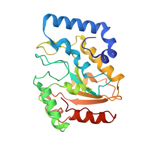
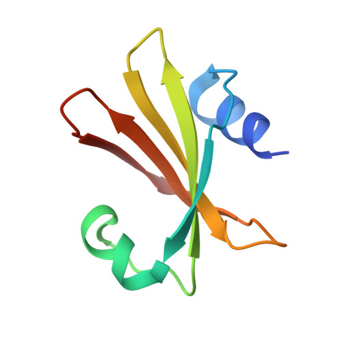
Structural and biophysical analysis of interactions between cod and human uracil-DNA N-glycosylase (UNG) and UNG inhibitor (Ugi).

Assefa, N.G.Niiranen, L.Johnson, K.A.Leiros, H.K.Smalas, A.O.Willassen, N.P.Moe, E.

(2014) Acta Crystallogr D Biol Crystallogr 70: 2093-2100

  • DOI: https://doi.org/10.1107/S1399004714011699
  • Primary Citation of Related Structures:  
    4LYL

  • PubMed Abstract: 

    Uracil-DNA N-glycosylase from Atlantic cod (cUNG) shows cold-adapted features such as high catalytic efficiency, a low temperature optimum for activity and reduced thermal stability compared with its mesophilic homologue human UNG (hUNG). In order to understand the role of the enzyme-substrate interaction related to the cold-adapted properties, the structure of cUNG in complex with a bacteriophage encoded natural UNG inhibitor (Ugi) has been determined. The interaction has also been analyzed by isothermal titration calorimetry (ITC). The crystal structure of cUNG-Ugi was determined to a resolution of 1.9 Å with eight complexes in the asymmetric unit related through noncrystallographic symmetry. A comparison of the cUNG-Ugi complex with previously determined structures of UNG-Ugi shows that they are very similar, and confirmed the nucleotide-mimicking properties of Ugi. Biophysically, the interaction between cUNG and Ugi is very strong and shows a binding constant (Kb) which is one order of magnitude larger than that for hUNG-Ugi. The binding of both cUNG and hUNG to Ugi was shown to be favoured by both enthalpic and entropic forces; however, the binding of cUNG to Ugi is mainly dominated by enthalpy, while the entropic term is dominant for hUNG. The observed differences in the binding properties may be explained by an overall greater positive electrostatic surface potential in the protein-Ugi interface of cUNG and the slightly more hydrophobic surface of hUNG.


  • Organizational Affiliation

    Department of Chemistry/Norstruct, UiT The Arctic University of Norway, 9037 Tromsø, Norway.


Macromolecules
Find similar proteins by:  (by identity cutoff)  |  3D Structure
Entity ID: 1
MoleculeChains Sequence LengthOrganismDetailsImage
Uracil-DNA glycosylase
A, C, E, G, I
A, C, E, G, I, K, M, O
223Gadus morhuaMutation(s): 0 
Gene Names: ung1
EC: 3.2.2.3
UniProt
Find proteins for Q9I983 (Gadus morhua)
Explore Q9I983 
Go to UniProtKB:  Q9I983
Entity Groups  
Sequence Clusters30% Identity50% Identity70% Identity90% Identity95% Identity100% Identity
UniProt GroupQ9I983
Sequence Annotations
Expand
  • Reference Sequence
Find similar proteins by:  (by identity cutoff)  |  3D Structure
Entity ID: 2
MoleculeChains Sequence LengthOrganismDetailsImage
Uracil-DNA glycosylase inhibitor
B, D, F, H, J
B, D, F, H, J, L, N, P
84Bacillus phage PBS2Mutation(s): 0 
Gene Names: UGI
UniProt
Find proteins for P14739 (Bacillus phage PBS2)
Explore P14739 
Go to UniProtKB:  P14739
Entity Groups  
Sequence Clusters30% Identity50% Identity70% Identity90% Identity95% Identity100% Identity
UniProt GroupP14739
Sequence Annotations
Expand
  • Reference Sequence
Experimental Data & Validation

Experimental Data

  • Method: X-RAY DIFFRACTION
  • Resolution: 1.93 Å
  • R-Value Free: 0.283 
  • R-Value Work: 0.237 
  • R-Value Observed: 0.239 
  • Space Group: P 1 21 1
Unit Cell:
Length ( Å )Angle ( ˚ )
a = 98.21α = 90
b = 86.92β = 90.35
c = 175.37γ = 90
Software Package:
Software NamePurpose
MOLREPphasing
REFMACrefinement
XDSdata reduction
XSCALEdata scaling

Structure Validation

View Full Validation Report


Entry History 

Deposition Data

Revision History  (Full details and data files)

  • Version 1.0: 2014-08-13
    Type: Initial release