4LUP

Crystal structure of the complex formed by region of E. coli sigmaE bound to its -10 element non template strand


Experimental Data Snapshot

  • Method: X-RAY DIFFRACTION
  • Resolution: 1.20 Å
  • R-Value Free: 0.165 
  • R-Value Work: 0.147 
  • R-Value Observed: 0.147 

wwPDB Validation   3D Report Full Report


This is version 1.3 of the entry. See complete history


Literature

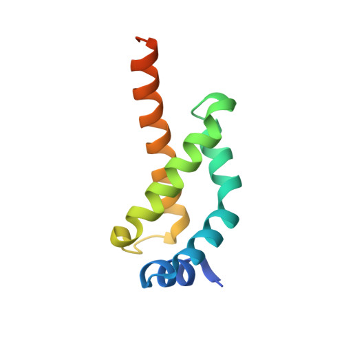
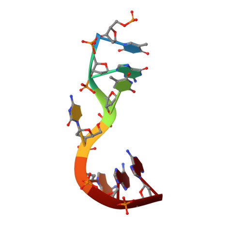
Structural basis for -10 promoter element melting by environmentally induced sigma factors.

Campagne, S.Marsh, M.E.Capitani, G.Vorholt, J.A.Allain, F.H.

(2014) Nat Struct Mol Biol 21: 269-276

  • DOI: https://doi.org/10.1038/nsmb.2777
  • Primary Citation of Related Structures:  
    2MAO, 2MAP, 4LUP

  • PubMed Abstract: 

    Bacterial transcription is controlled by sigma factors, the RNA polymerase subunits that act as initiation factors. Although a single housekeeping sigma factor enables transcription from thousands of promoters, environmentally induced sigma factors redirect gene expression toward small regulons to carry out focused responses. Using structural and functional analyses, we determined the molecular basis of -10 promoter element recognition by Escherichia coli σ(E), which revealed an unprecedented way to achieve promoter melting. Group IV sigma factors induced strand separation at the -10 element by flipping out a single nucleotide from the nontemplate-strand DNA base stack. Unambiguous selection of this critical base was driven by a dynamic protein loop, which can be substituted to modify specificity of promoter recognition. This mechanism of promoter melting explains the increased promoter-selection stringency of environmentally induced sigma factors.


  • Organizational Affiliation

    1] Institute of Microbiology, Eidgenössische Technische Hochschule (ETH) Zurich, Zurich, Switzerland. [2] Institute of Molecular Biology and Biophysics, Eidgenössische Technische Hochschule (ETH) Zurich, Zurich, Switzerland.


Macromolecules

Find similar proteins by:  (by identity cutoff)  |  3D Structure
Entity ID: 1
MoleculeChains Sequence LengthOrganismDetailsImage
RNA polymerase sigma factorA,
B [auth C]
106Escherichia coliMutation(s): 0 
Gene Names: rpoEBN17_18601ECs3439LF82_1962
UniProt
Find proteins for Q0P6M2 (Escherichia coli)
Explore Q0P6M2 
Go to UniProtKB:  Q0P6M2
Entity Groups  
Sequence Clusters30% Identity50% Identity70% Identity90% Identity95% Identity100% Identity
UniProt GroupQ0P6M2
Sequence Annotations
Expand
  • Reference Sequence

Find similar nucleic acids by:  Sequence   |   3D Structure  

Entity ID: 2
MoleculeChains LengthOrganismImage
region 2 of sigmaE of E. coliC [auth B],
D
7N/A
Sequence Annotations
Expand
  • Reference Sequence
Small Molecules
Ligands 1 Unique
IDChains Name / Formula / InChI Key2D Diagram3D Interactions
EDO
Query on EDO

Download Ideal Coordinates CCD File 
E [auth A]1,2-ETHANEDIOL
C2 H6 O2
LYCAIKOWRPUZTN-UHFFFAOYSA-N
Experimental Data & Validation

Experimental Data

  • Method: X-RAY DIFFRACTION
  • Resolution: 1.20 Å
  • R-Value Free: 0.165 
  • R-Value Work: 0.147 
  • R-Value Observed: 0.147 
  • Space Group: P 1 21 1
Unit Cell:
Length ( Å )Angle ( ˚ )
a = 39.05α = 90
b = 36.48β = 92.48
c = 73.37γ = 90
Software Package:
Software NamePurpose
XSCALEdata scaling
PHENIXrefinement
PDB_EXTRACTdata extraction
DA+data collection
XDSdata reduction
PHASERphasing

Structure Validation

View Full Validation Report


Entry History 

Deposition Data

Revision History  (Full details and data files)

  • Version 1.0: 2014-02-19
    Type: Initial release
  • Version 1.1: 2014-02-26
    Changes: Database references
  • Version 1.2: 2014-03-19
    Changes: Database references
  • Version 1.3: 2023-09-20
    Changes: Data collection, Database references, Derived calculations, Refinement description