4DYG

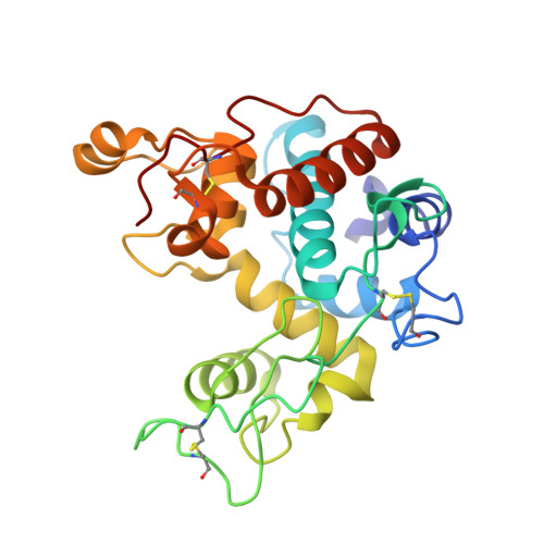
Crystal Structure of a Family GH-19 Chitinase from rye seeds in complex with (GlcNAc)4


Experimental Data Snapshot

  • Method: X-RAY DIFFRACTION
  • Resolution: 1.70 Å
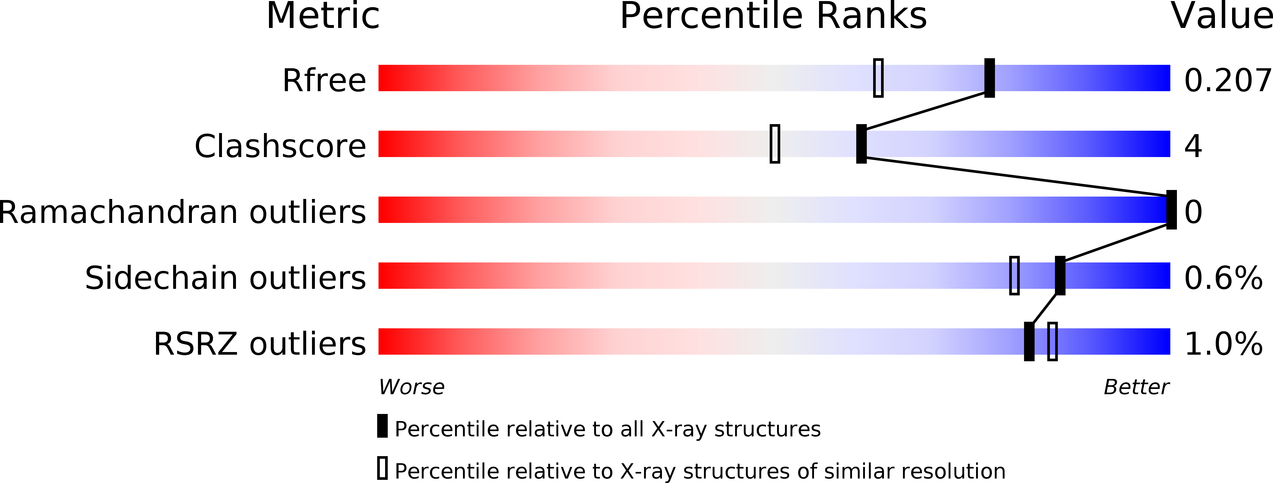
  • R-Value Free: 0.212 
  • R-Value Work: 0.183 
  • R-Value Observed: 0.183 

wwPDB Validation   3D Report Full Report


This is version 2.1 of the entry. See complete history


Literature

Crystal structure and chitin oligosaccharide-binding mode of a 'loopful' family GH19 chitinase from rye, Secale cereale, seeds

Ohnuma, T.Numata, T.Osawa, T.Inanaga, H.Okazaki, Y.Shinya, S.Kondo, K.Fukuda, T.Fukamizo, T.

(2012) FEBS J 279: 3639-3651

  • DOI: https://doi.org/10.1111/j.1742-4658.2012.08723.x
  • Primary Citation of Related Structures:  
    4DWX, 4DYG

  • PubMed Abstract: 

    The substrate-binding mode of a 26-kDa GH19 chitinase from rye, Secale cereale, seeds (RSC-c) was investigated by crystallography, site-directed mutagenesis and NMR spectroscopy. The crystal structure of RSC-c in a complex with an N-acetylglucosamine tetramer, (GlcNAc)(4) , was successfully solved, and revealed the binding mode of the tetramer to be an aglycon-binding site, subsites +1, +2, +3, and +4. These are the first crystallographic data showing the oligosaccharide-binding mode of a family GH19 chitinase. From HPLC analysis of the enzymatic reaction products, mutation of Trp72 to alanine was found to affect the product distribution obtained from the substrate, p-nitrophenyl penta-N-acetyl-β-chitopentaoside. Mutational experiments confirmed the crystallographic finding that the Trp72 side chain interacts with the +4 moiety of the bound substrate. To further confirm the crystallographic data, binding experiments were also conducted in solution using NMR spectroscopy. Several signals in the (1) H-(15) N HSQC spectrum of the stable isotope-labeled RSC-c were affected upon addition of (GlcNAc)(4) . Signal assignments revealed that most signals responsive to the addition of (GlcNAc)(4) are derived from amino acids located at the surface of the aglycon-binding site. The binding mode deduced from NMR binding experiments in solution was consistent with that from the crystal structure.


  • Organizational Affiliation

     Department of Advanced Bioscience, Kinki University, Nara, Japan Biomedical Research Institute, National Institute of Advanced Industrial Science and Technology (AIST), Tsukuba, Japan.


Macromolecules
Find similar proteins by:  (by identity cutoff)  |  3D Structure
Entity ID: 1
MoleculeChains Sequence LengthOrganismDetailsImage
Basic endochitinase C
A, B
244Secale cerealeMutation(s): 0 
Gene Names: rscc
EC: 3.2.1.14
UniProt
Find proteins for Q9FRV0 (Secale cereale)
Explore Q9FRV0 
Go to UniProtKB:  Q9FRV0
Entity Groups  
Sequence Clusters30% Identity50% Identity70% Identity90% Identity95% Identity100% Identity
UniProt GroupQ9FRV0
Sequence Annotations
Expand
  • Reference Sequence
Oligosaccharides

Help

Entity ID: 2
MoleculeChains Length2D Diagram Glycosylation3D Interactions
2-acetamido-2-deoxy-beta-D-glucopyranose-(1-4)-2-acetamido-2-deoxy-beta-D-glucopyranose-(1-4)-2-acetamido-2-deoxy-beta-D-glucopyranose-(1-4)-2-acetamido-2-deoxy-beta-D-glucopyranose
C, D
4N/A
Glycosylation Resources
GlyTouCan:  G01361SX
GlyCosmos:  G01361SX
GlyGen:  G01361SX
Small Molecules
Ligands 3 Unique
IDChains Name / Formula / InChI Key2D Diagram3D Interactions
MES
Query on MES

Download Ideal Coordinates CCD File 
E [auth A],
L [auth B]
2-(N-MORPHOLINO)-ETHANESULFONIC ACID
C6 H13 N O4 S
SXGZJKUKBWWHRA-UHFFFAOYSA-N
SO4
Query on SO4

Download Ideal Coordinates CCD File 
F [auth A],
G [auth A],
M [auth B]
SULFATE ION
O4 S
QAOWNCQODCNURD-UHFFFAOYSA-L
ZN
Query on ZN

Download Ideal Coordinates CCD File 
H [auth A],
I [auth A],
J [auth A],
K [auth A],
N [auth B]
ZINC ION
Zn
PTFCDOFLOPIGGS-UHFFFAOYSA-N
Experimental Data & Validation

Experimental Data

  • Method: X-RAY DIFFRACTION
  • Resolution: 1.70 Å
  • R-Value Free: 0.212 
  • R-Value Work: 0.183 
  • R-Value Observed: 0.183 
  • Space Group: P 21 21 21
Unit Cell:
Length ( Å )Angle ( ˚ )
a = 64.695α = 90
b = 85.387β = 90
c = 108.364γ = 90
Software Package:
Software NamePurpose
HKL-2000data collection
CNSrefinement
HKL-2000data reduction
HKL-2000data scaling
CNSphasing

Structure Validation

View Full Validation Report


Entry History 

Deposition Data

Revision History  (Full details and data files)

  • Version 1.0: 2012-08-15
    Type: Initial release
  • Version 1.1: 2013-06-26
    Changes: Database references
  • Version 2.0: 2020-07-29
    Type: Remediation
    Reason: Carbohydrate remediation
    Changes: Atomic model, Data collection, Database references, Derived calculations, Structure summary
  • Version 2.1: 2023-11-08
    Changes: Data collection, Database references, Refinement description, Structure summary