The two faces of the Escherichia coli 23 S rRNA sarcin/ricin domain: the structure at 1.11 A resolution.
Correll, C.C., Wool, I.G., Munishkin, A.(1999) J Mol Biol 292: 275-287
- PubMed: 10493875
- DOI: https://doi.org/10.1006/jmbi.1999.3072
- Primary Citation of Related Structures:
480D, 483D - PubMed Abstract:
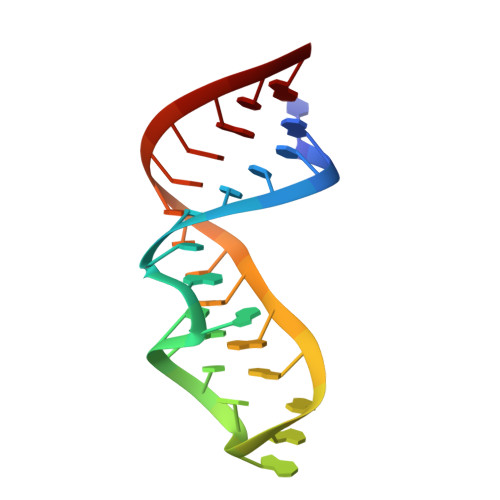
The sarcin/ricin domain of 23 S - 28 S ribosomal RNA is essential for protein synthesis because it forms a critical part of the binding site for elongation factors. A crystal structure of an RNA of 27 nucleotides that mimics the domain in Escherichia coli 23 S rRNA was determined at 1.11 A resolution. The domain folds into a hairpin distorted by four non-canonical base-pairs and one base triple. The fold is stabilized by cross-strand and intra-stand stacking; no intramolecular stabilizing metal ions are observed. This is the first structure to reveal in great detail the geometry and the hydration of two common motifs that are conserved in this rRNA domain, a GAGA tetraloop and a G-bulged cross-strand A stack. Differences in the region connecting these motifs to the stem in the E. coli and in the rat sarcin/ricin domains may contribute to the species-specific binding of elongation factors. Correlation of nucleotide protection data with the structure indicates that the domain has two surfaces. One surface is accessible, lies primarily in the major groove, and is likely to bind the elongation factors. The second lies primarily in the minor groove, and is likely to be buried in the ribosome. This minor groove surface includes the Watson-Crick faces of the cytosine bases in the unusual A2654.C2666 and U2653.C2667 water-mediated base-pairs.
Organizational Affiliation:
Department of Biochemistry and Molecular Biology, The University of Chicago, Chicago, IL 60637, USA. ccorrell@midway.uchicago.edu